This is an automatically translated article.
Article written by MSc, Dr. Dang Manh Cuong, Department of Diagnostic Imaging - Vinmec Central Park International General Hospital
Small intestine magnetic resonance is a special imaging method that helps doctors observe the structure of small intestine loops, thereby accurately diagnosing diseases such as small bowel abscess, intestinal obstruction, complications of Crohn's disease and other inflammatory bowel diseases.
1. What is small intestine magnetic resonance (MRI)?
Magnetic resonance small intestine (MR enterography) is a special imaging method that uses magnetic resonance (MRI) with contrast material to create detailed images of the small intestine to help doctors evaluate conditions. diseases of the small intestine.
This method uses a magnetic field to create detailed images of your body's organs. The computer then analyzes the images. Prior to examination, the patient will be given intravenous contrast and contrast agents to highlight the small intestine. A medication will also be injected to decrease bowel movements to increase image quality.
This is not an X-ray imaging technique and it does not involve any radiation. Oral contrast also does not contain any radioactive material. Small intestine MRI scans give a fairly detailed picture of the structure of the small bowel loops. The exam can take about 45 minutes.
2. Why use small intestine magnetic resonance?
This method is used to:
Diagnose the presence and complications of Crohn's disease and other inflammatory bowel diseases; Identify the source of bleeding and vascular abnormalities of the small intestine; Small bowel tumor ; Abscesses and fistulas; Bowel obstruction . Especially in Crohn's disease, it is often recommended when Crohn's disease is suspected because Crohn's disease tends to strike young people, who are more at risk of problems from high radiation exposure. time. This method can help avoid unnecessary X-rays. In addition, this method is also a better method of evaluating soft tissue problems than other methods.

3. Is there any risk of small bowel MRI?
The magnetic field of an MRI machine can change the behavior of any medical device implanted in the body. Magnetic contrast agents can cause fibrosis of the renal system if your kidney function is not working properly. Some people have an allergic reaction to the contrast agent, although it's very rare. There may be other risks, depending on your specific medical condition. As such, be sure to discuss any concerns with your doctor prior to having the scan performed.
4. What do you need to prepare before having an MRI of the small intestine?
Before having this procedure, you may need to:
Have any blood tests or other tests ordered by the hospital where you have the scan done. Tell your doctor or technician if you are or may be pregnant. Inform your doctor or technician if you have or use any implanted medical device, such as a hearing aid... If you have such devices, you should not perform this procedure. it's France. For example, if you have a defibrillator, pacemaker, cochlear implant, blood vessel clamp in a brain aneurysm... you should not perform this examination or enter the MRI area unless your radiologist says it's doable. . Ask your doctor if you should stop any medications you're taking. Ask your doctor when to stop eating and drinking. You may be asked to avoid certain foods or drinks, such as carbonated drinks. You may also be asked not to eat or drink for 6 hours before the search is performed. Tell your doctor about any allergies or other health conditions you have, such as diabetes or kidney disease. Tell your doctor about whether you may need a sedative to relax during the test. Do not wear any jewelry on your body, or bring any personal items with you when performing the search. Do not bring any metal objects into the studio, including hairpins and metal zippers. If you have sensitive hearing, ask to wear earplugs during the procedure. The MRI machine can make a loud noise that some people may find uncomfortable. If you are able to go home the same day, in case you are sedated before the procedure, make sure you have someone drive you home.
5. How is the process done?
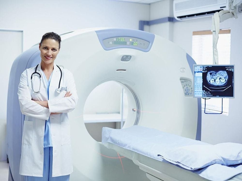
You will be changed into a hospital gown before entering the imaging room; You will be given a contrast medium before the scan is performed. The scan will begin about an hour after you drink the contrast; The technician will help stabilize and position you on the table in the imaging room. The more still you lie down, the better and clearer the image will be; The nurse or technician will inject contrast medicine from a vein during the scan; An MRI machine will take pictures before and after the contrast is injected intravenously. You will be alone in the room, but you will still be able to talk to people outside the room through the speakers. The camera may make some noise during shooting. This is normal; If during the scan you will be asked to hold your breath for short periods of time, try to hold your breath to get a good picture; You need to stay still during the shoot.
6. Problems after shooting
Some people have mild nausea, cramps, or diarrhea from the contrast agent or other drugs being injected into the body. Inform your doctor if you have any discomfort in your body.

7. Some limitations of small bowel MRI
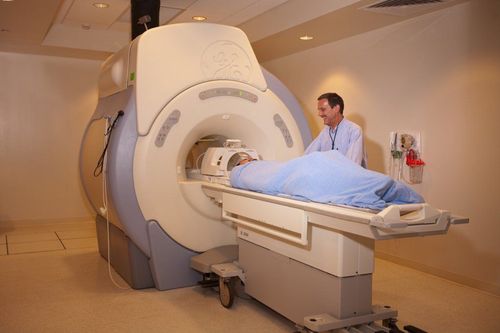
Wanting high quality images depends on your ability to lie still and follow the instructions to hold your breath while shooting. If you are nervous, confused, or in severe pain, you may find it difficult to lie still while shooting, thereby affecting image quality. An oversized person may not be suitable for some types of MRI machines. Implants and other metal objects in the body can make it difficult to get clear images. Patient movements can have a similar effect. Very irregular heart rhythms can affect image quality. This is because some imaging techniques are based on the electrical activity of the heart. Although there is no evidence that MRI harms an unborn baby, pregnant women should not have an MRI during the first trimester unless it is medically necessary. For optimal results, the patient should receive the full amount of oral contrast, lie still, and follow the technician's breathing instructions. Furthermore, results may be affected if the patient is unable to use intravenous contrast agent (gadolinium). Vinmec International General Hospital put into use the 3.0 Tesla Silent technology magnetic resonance imaging machine. Magnetic resonance imaging machine 3.0 Tesla with Silent technology of GE Healthcare (USA).
Silent technology is especially beneficial for patients who are children, the elderly, weak health patients and patients undergoing surgery. Limiting noise, creating comfort and reducing stress for customers during the shooting process, helping to capture better quality images and shorten the shooting time. Magnetic resonance imaging technology is the technology applied in the most popular and safest imaging method today because of its accuracy, non-invasiveness and non-X-ray use.
Please dial HOTLINE for more information or register for an appointment HERE. Download MyVinmec app to make appointments faster and to manage your bookings easily.


