This is an automatically translated article.
The article was professionally consulted by Dr. Nguyen Anh Tu - Doctor of Obstetric Ultrasound - Prenatal Diagnosis - Obstetrics Department - Vinmec Hai Phong International General Hospital.Morphological ultrasound is a technique used to monitor the formation and development of the fetus in the womb, this technique can be performed between the 20th and 24th weeks of pregnancy.
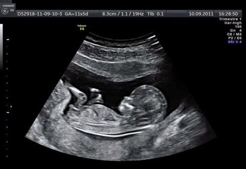
1. What is morphological ultrasound technique?
Morphological ultrasound is an ultrasound technique that allows to observe the image of the fetus in the mother's womb, including the external appearance and internal organs, thereby helping to monitor the formation and fetal development, and detect some abnormalities (if any).

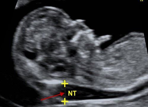
Siêu âm hình thái học giúp phát hiện dị tật thai nhi (nếu có)
2. Meaning of morphological ultrasound technique
Ultrasound in general and morphological ultrasound in particular have great significance in each periodical antenatal check-up, specifically:
Monitor and record fetal movements and fetal morphology in the womb. Detect any abnormalities if any of the fetus. Estimated date of birth.
Trắc nghiệm: Xét nghiệm Triple test là gì? Cần thực hiện khi nào?
Triple test và một trong những xét nghiệm sàng lọc trước sinh quan trọng nhất trong thai kỳ, giúp chẩn đoán nguy cơ dị tật thai nhi, là cơ sở để các bác sĩ chỉ định thực hiện các xét nghiệm sàng lọc xâm lấn như chọc ối, sinh thiết gai nhau. Theo dõi bài viết sau để biết Triple test là gì và nên thực hiện khi nào?3. How many weeks can pregnant women perform morphological ultrasound?
Fetal morphology ultrasound can be done between 20-24 weeks of pregnancy. This is considered a golden time and is mandatory to evaluate the structure and morphology of the fetus, screen for abnormalities if any.
At this time, morphological ultrasound allows to observe external morphology, internal organ structure. Ultrasound also allows the fetal heart to be heard. Through biological indicators and size, doctors can make an assessment of whether the fetus is developing normally according to gestational age. Compared to the following weeks of pregnancy, this is a favorable time because the fetus has developed and perfected internal and external organs. Ultrasound also shows abnormalities, if any, from which to advise and guide pregnant women to monitor and provide appropriate and best pregnancy care.

20 -24 tuần tuổi được xem là thời điểm vàng để siêu âm hình thái thai nhi
4. Morphological ultrasound results
When performing morphological ultrasound in the period from 20-24 weeks, the doctor will in turn check from the top to the bottom of the feet, from the outside to the inside to fully diagnose the development of the fetus. The results of morphological ultrasound include the following observations and indicators:
Head: Image of skull arch bone, measurement of head circumference and diameter, brain structure, fully developed facial part 2 eye sockets, nose, mouth and chin. Ultrasound helps to diagnose anomalies of the head and face, if present, including morphological and structural abnormalities of the brain, cleft lip, cleft palate, nasopharynx, and cleft palate tumors. Spine: Morphological ultrasound at 22 weeks of pregnancy allows to check for abnormalities in the spine such as spondylolisthesis, open vertebral split. Chest: Image and position, size of the fetal heart as lying on the left side of the chest, with 4 chambers with 2 upper atria, 2 lower ventricles. Picture of major blood vessels in the chest including the aorta and pulmonary artery. Ultrasound helps diagnose abnormalities if present in the heart and lungs including abnormalities in the size of the heart chambers, aorta, pulmonary artery, heart tumor, total dilated heart, pleural effusion,. .. Abdomen: Morphological ultrasound shows images and structures of the diaphragm, stomach, kidneys and bladder, and measurements of the abdomen. Ultrasound helps diagnose abnormalities such as esophageal atresia, duodenal obstruction, small bowel obstruction, colonic obstruction, anorectal malformation, umbilical hernia, abdominal wall cleft, bladder malformation, no kidney, pyelonephritis, polycystic kidney, ... Hands and feet: Image of extremities including 2 hands, 2 legs with full 5 fingers, 5 toes and 3 segments in each limb, right foot square angle to the shin, the measure of the length of the femur. Ultrasound helps diagnose abnormalities if present in limbs such as short, short, crooked, missing or extra fingers on hands and feet. In addition to the morphology and structure of the organs and organs mentioned above, morphological ultrasound between 20 - 24 weeks of pregnancy also evaluates the appendages including:
Place of placenta: Location of placenta low or high (before or after the 20th week of pregnancy). Ultrasound helps diagnose abnormalities in the placenta such as placental tumor, placenta previa, placenta previa, thick placenta, low placenta, ... Umbilical cord: The umbilical cord is full of 3 blood vessels including 2 arteries. and 1 vein. Amniotic fluid: Ultrasound evaluates the color and amount of amniotic fluid through the amniotic index. Ultrasound also helps diagnose abnormalities in amniotic fluid such as oligohydramnios, polyhydramnios, and amniotic septum. Morphological ultrasound plays an important role in checking pregnancy health, helping to diagnose and detect abnormalities and abnormalities in fetal development.
At Vinmec International General Hospital, there is a package maternity service as a solution to help pregnant women feel secure because of the companionship of the medical team throughout the pregnancy. When choosing Maternity Package, pregnant women can:
The pregnancy process is monitored by a team of qualified doctors Regular check-up, early detection of abnormalities Maternity package helps to facilitate the process. Birth program Newborns receive comprehensive care To register for examination and treatment at Vinmec International General Hospital, you can contact Vinmec Health System nationwide, or register online HERE HERE
SEE MORE
The indicators in the fetal ultrasound results at 32 weeks, how many kg is standard? The process of fetal formation and development week by week