This is an automatically translated article.
Whether the fetal ultrasound can detect the congenital heart is the concern of many pregnant women. Currently, with the advancement of medicine, fetal echocardiography can help detect and diagnose congenital heart defects in the fetus.
1. Congenital heart disease in the fetus
Congenital heart defects in the fetus are one of the most common abnormalities and the leading cause of death in newborns. Most cases of babies born with congenital heart disease are missed or undiagnosed during pregnancy. Congenital heart greatly affects the health and comprehensive development of children after birth. Most children with congenital heart disease need surgery right after birth, in severe cases, children need emergency resuscitation and treatment with the cooperation of doctors from many specialties. Without surgery, the baby can face poor health after birth, dangerously leading to death.

Dị tật tim bẩm sinh ảnh hưởng lớn tới sức khỏe và sự phát triển của trẻ sau này
2. Can fetal ultrasound detect a congenital heart?
Currently, modern medicine has helped detect and diagnose most congenital heart defects in the fetus such as aortic stenosis, arrhythmia, ....
Diagnosis and early detection of heart Congenital births in pregnancy play an extremely important role in identifying malformations, providing timely intervention solutions, thereby, improving and improving the health and life chances of children after birth. Except for cases of severe congenital heart disease and poor prognosis, doctors will appoint pregnant women to terminate the pregnancy.
3. Which cases need fetal echocardiography?
In the following cases, it is necessary to perform fetal echocardiography to screen and diagnose heart defects:
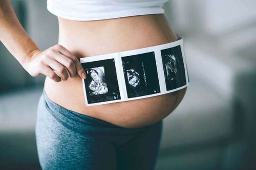
Cases originating from the mother: The mother has a congenital heart defect; suffering from metabolic disorders such as diabetes mellitus, Phenylketonuria, lupus erythematosus, ...; been exposed to many stimulants such as alcohol, beer, tobacco, ...; taking biosynthetic inhibitors such as ibuprofen, salicylic acid, or antiepileptic drugs...; infected with viruses such as Rubella virus, Coxsackie, Parvovirus, ...; mothers using in vitro fertilization. Cases originating from the fetus: ultrasound suspected congenital heart in the fetus; fetal growth retardation, short femur length,... ; other malformations such as umbilical hernia , esophageal atrophy , diaphragmatic hernia , ... ; chromosomal abnormalities; twin-twin transfusion syndrome, multiple pregnancy; have an arrhythmia; pregnancy edema; nuchal translucency > 3mm (done at 10-12 weeks of pregnancy); ... From 12 weeks of age, the fetus has developed relatively fully in terms of morphology and has reflexes such as flexing and stretching the body, stretching the limbs... This is also one of the three abnormal ultrasound landmarks. Important defects recommended by experts to be done. During this ultrasound, doctors will especially check and screen for early abnormalities of the brain, face, heart, digestive, urinary, extremities and the whole body. Because the baby is still quite small, a high-end 4D ultrasound system like the GE Voluson E10 will play a very important role in helping doctors at Vinmec detect more than 95% of abnormalities during this period. Packed with the latest technologies, the GE Voluson E10 enables enhanced image quality and penetration for outstanding high-resolution images and easy operation. Cases of family origin: Children before or in previous pregnancies with congenital heart defects; father with congenital heart disease; a history of an inherited genetic disorder; ...

Nếu gia đình có trường hợp bị tim bẩm sinh thai phụ cần siêu âm tim thai
4. How is fetal echocardiography done, at what time?
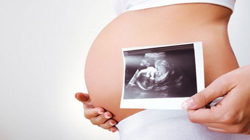
Fetal echocardiography for the diagnosis, detection and screening of congenital heart disease is performed between 18-24 weeks of pregnancy. Doctors will apply 4-dimensional (4D) ultrasound to survey the structure of the fetal cardiovascular system, the imaging results can detect most congenital heart defects from mild to severe.
Fetal echocardiography can detect congenital heart, but this test is often missed in prenatal examination, affecting the development and health of the baby after birth.
At Vinmec International General Hospital, there is a package maternity service as a solution to help pregnant women feel secure because of the companionship of the medical team throughout the pregnancy. When choosing Maternity Package, pregnant women can:
The pregnancy process is monitored by a team of qualified doctors Regular check-up, early detection of abnormalities Maternity package helps to facilitate the process. birthing process Newborns get comprehensive care
Please dial HOTLINE for more information or register for an appointment HERE. Download MyVinmec app to make appointments faster and to manage your bookings easily.
Recommended video:
12 week old fetus clearly seen from 4d fetal ultrasound