This is an automatically translated article.
Articles written by MSc, BS. Dang Manh Cuong, Department of Diagnostic Imaging, Vinmec Central Park International General Hospital
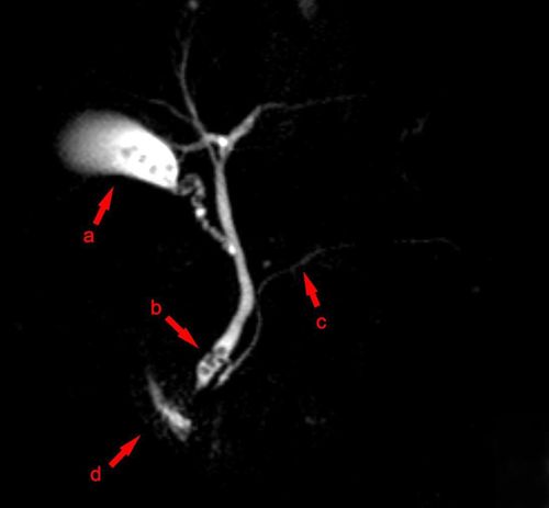
Magnetic resonance imaging (MRI) is the most valuable imaging modality in the evaluation of spinal pathologies. Magnetic resonance imaging of the lumbar spine helps to evaluate and detect many diseases in the lumbar spine and is used to monitor changes after surgery.
1. What is magnetic resonance?
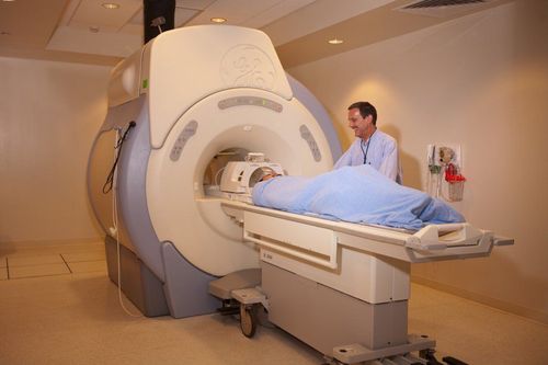
Magnetic resonance imaging (MRI) is a non-invasive, non-radiative (X-ray) imaging modality used to diagnose medical conditions. An MRI uses a strong magnetic field, radio waves, and a computer to create detailed images of structures inside the body.
MRI images allow doctors to examine the inside of the body and detect medical conditions. The resulting images can be viewed on a computer monitor. These images can also be sent electronically, printed or copied to CD, DVD, or uploaded to the digital cloud.

Chụp cộng hưởng từ (MRI) cho phép chẩn đoán hình ảnh chính xác, an toàn
2. Purpose of lumbar spine MRI
Currently, MRI is the most valuable imaging modality in the evaluation of spinal pathologies.
MRI is used to evaluate or detect:
Spine anatomical structure and spinal curve abnormality or not; Birth defects of the vertebrae or spinal cord; Traumatic injuries to bones, discs, ligaments or spinal cord; Disc and joint diseases. Both are common causes of severe low back pain and sciatica (back pain that radiates to the lower extremities); Compression or inflammation of the spinal cord and nerve roots; Infection of the vertebrae, discs, spinal cord, or dura; Tumors in the vertebrae, spinal cord, nerves, or surrounding soft tissues. Lumbar spine MRI is also used to help plan surgery such as decompression of pinched nerves, spinal fusion, or steroid injections. Steroid injections to relieve pain are usually done under X-ray guidance.
MRI of the spine can detect other causes of back pain such as depression or bone marrow edema. It is also used to monitor post-operative changes (scarring or infection).

Người bệnh bị đau thắt lưng được chỉ định chụp MRI
3. Who gets an MRI of the lumbar spine?
All people suspected of having lumbar spine disease can be ordered to be scanned, except for some cases with absolute contraindications not to enter the imaging room.4. What does the patient need to prepare?
Patient needs to change into hospital clothes before the scan or his or her own clothes if no metal tools are inside; Patients can eat and drink as usual or may be instructed on eating and drinking before performing MRI depending on each specific indication; If MRI with contrast agent injection is indicated. You may be asked if you have asthma or are allergic to iodine contrast agents, drugs, foods or other allergic factors because the contrast agent used is Gadolinium. Gadolinium may be used in patients with an iodine contrast allergy. Gadolinium is less prone to allergic reactions than iodinated contrast agents. However, even if the patient is allergic to gadolinium, it can be used after appropriate anti-allergy medication prior to the scan; The patient should tell the technician or the chiropractor about any abnormalities, if any, or recent surgery; Because during an MRI scan requires absolute rest, infants and young children often need sedation or anesthesia before the scan. Therefore, the anesthesiologist will administer anesthesia during the scan to prevent the patient's movements from interfering with the image. You will be told how to prepare your child;

Thuốc an thần có thể được sử dụng đối với những trường hợp kích động
Leave all jewelry and other metal accessories at home or remove them before the MRI because metal and electronic items can interfere with the magnetic field of the MRI machine, damaging the device, causing burns or interfere with the captured images, so do not bring them into the shooting room. Things not to bring during an MRI: + Jewelry, watches, credit cards and hearing aids, cell phones, electronic watches and monitoring devices. These items can be damaged when entering the MRI machine room.
+ Pins, hairpins, metal zippers and similar metal tools. These items can distort the MRI image.
+ Removable dentures;
+ Pen, pocket knife and eyeglasses.
In most cases, an MRI is safe for patients with metal implants, with some exceptions. People with the following implants may not and should not enter the MRI area without first being evaluated for safety: + Certain cochlear (ear) implants;
+ Certain metals are used for brain aneurysms;
+ Several metal coils are placed in the blood vessels;
+ Some defibrillators and pacemakers.
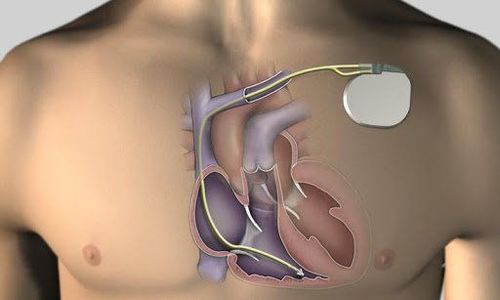
Bệnh nhân có máy tạo nhịp tim không thể chụp MRI
Notify technician or radiologist if patient has shrapnel, bullets, or other metal that may be present in the body. Foreign bodies near and especially in the orbit are important because they can move or heat up during the scan and can cause blindness. The inks used in tattoos may contain iron and thus can become hot during the MRI scan, causing skin burns (this is rare). Fillings, braces, eyeshadows and some other cosmetics are not usually affected by magnetic fields, but they can distort images in the face or brain area. Anyone accompanying a patient into the MRI room should be checked for metal or implanted devices.
5. What is the shooting process like?
The patient lies on the table, fixed with a strap, helping the patient stay still and immobilize the position. A device (called a coil capable of transmitting and receiving radio waves) is installed around the part to be photographed. The imaging process typically involves multiple pulses and echoes, some of which can last for several minutes. Gadolinium contrast agents are used when evaluating for infections, tumors, or recurrent disc problems after surgery. If magnetic contrast material is used, the doctor, nurse, or technician will place a needle into a vein in your arm before the scan that is used to inject contrast. The patient is inserted into the large magnet block of the MRI machine. The technician will perform the scan with an imaging control system in the control room outside the MRI machine room. Once the scan is over, you may be asked to wait while the radiologist checks that the images have passed. If you pass the test, you will be taken out of the room to complete the scan. The scan is usually completed in about 30 minutes. If magnetic contrast material is used, approximately 15 minutes will be added to the total scan time.
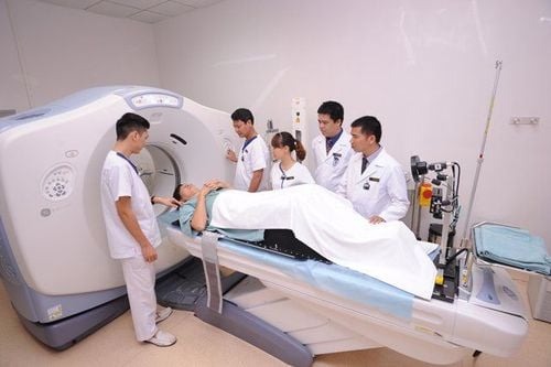
Kỹ thuật viên sẽ hướng dẫn bệnh nhân tư thế và lưu ý khi chụp MRI
6. How does it feel while shooting?
Most lumbar spine MRI scans are not uncomfortable. However, some patients find it uncomfortable to lie still for long periods of time, while others may feel claustrophobic while lying on the MRI machine. Some feel the noise from the MRI machine. The patient may feel warmth while lying in the imaging area of the MRI machine. The patient may experience noise from the scanner. Therefore, the patient will be provided with earplugs or headphones to reduce the noise generated by the scanner. Between pulses, the patient may feel quiet, so the patient can relax during this time. However, the patient must remain in the same position without moving. The patient was alone in the MRI room. However, the technician will be able to see, hear and talk to the patient at all times via the communication device on the machine. Some hospitals may allow a loved one into the room if they have also been safely screened. If the patient does not require sedation, there is no need for a post-sedation follow-up period. Patients can resume normal activities and diet immediately after the scan. In very rare cases, a few patients experience side effects from the contrast agent. These may include nausea, headache, and pain at the injection site. Very rarely, patients have had a rash or other allergic reaction to the contrast agent. If you have symptoms of an allergy, tell the technologist so the radiologist or other doctor can help right away.

Một số bệnh nhân mắc chứng sợ không gian kín có thể thấy khó chịu trong khi chụp MRI
7. Advantages and disadvantages of lumbar spine MRI
7.1 Advantages
No radiation exposure; MRI images of the spine are clearer and more detailed than those obtained with other imaging methods. MRI reveals spinal abnormalities, injuries, and pathologies that may not be detected by other imaging methods. MRI is the best method available to evaluate the spinal cord and nerve roots. MRI can detect abnormalities that are obscured by bone using other imaging methods. Gadolinium contrast agents are less likely to cause allergic reactions than iodinated contrast agents used in X-rays and CT scans. Useful in the evaluation of spinal cord injuries. It helps diagnose or rule out acute spinal cord compression when physical examination reveals muscle weakness or paralysis. MRI is the best method to evaluate ligament damage due to trauma. An MRI can detect early signs of an infection or tumor. MRI is more sensitive than CT for evaluating tumors, abscesses, and other soft tissue masses near the spinal cord. MRI is the preferred method for evaluating potential complications of surgery, including bleeding, scarring, infection, and recurrent disc herniation.

Chụp MRI cột sống thắt lưng giúp phát hiện dấu hiệu sớm của nhiễm trùng hoặc khối u
7.2 Cons
Lumbar spine MRI poses no risk to the patient as long as safety guidelines are followed. If sedatives are used, there is a risk of using too much. Therefore, the patient will be closely monitored during the scan to minimize this risk. The high magnetic field is not harmful. However, it can cause implanted devices in the body to malfunction or cause image distortion. Systemic renal fibrosis is a recognized, but rare, complication associated with Gadolinium contrast injection. It usually occurs in patients with severe kidney disease. The doctor will evaluate the patient's kidney function before considering if contrast can be injected. The risk of an allergic reaction is very slight if contrast material is used. Such allergic reactions are usually mild and controlled with medication. If the patient has an allergic reaction, the doctor will assist in handling it immediately. The manufacturers of intravenous contrast agents say that mothers should not breastfeed for 24 hours after using the contrast agent. However, the most recent ACR guidelines for magnetic contrast agents are studies showing that the amount of magnetic contrast absorbed by infants during breastfeeding is very low.

Sau khi chụp MRI có sử dụng chất tương phản không nên cho con bú trong 24 giờ
8. What are the limitations of spinal MRI?
The image will be shaky or blurry if you hold your breath poorly or you move around during shooting. Patients with large weight or size may not fit the MRI cage. Implants and other metal objects can cause interference that causes images to be distorted or unclear. Patient movements can have a similar effect. Irregular heart rhythms can affect image quality because some MRI techniques rely on the electrical activity of the heart. MRI is generally not recommended in critically injured patients because immobilizers and life support devices can distort MR images. However, the decision to have a scan or not is based on clinical judgment. In some trauma patients an MRI may also be required. Although there is no reason to believe that an MRI is safe for the fetus, however, women in the first trimester should not have an MRI unless it is medically necessary. Vinmec International General Hospital put into use the 3.0 Tesla Silent technology magnetic resonance imaging machine. Magnetic resonance imaging machine 3.0 Tesla with Silent technology of GE Healthcare (USA).
Silent technology is especially beneficial for patients who are children, the elderly, weak health patients and patients undergoing surgery. Limiting noise, creating comfort and reducing stress for customers during the shooting process, helping to capture better quality images and shorten the shooting time. Magnetic resonance imaging technology is the technology applied in the most popular and safest imaging method today because of its accuracy, non-invasiveness and no X-ray. About health you should visit and consult with specialists.
Reference source: radiologyinfo.org
Please dial HOTLINE for more information or register for an appointment HERE. Download MyVinmec app to make appointments faster and to manage your bookings easily.