This is an automatically translated article.
The article is professionally consulted by Master, Doctor Do Xuan Chien and Master, Doctor Hoang Thi Hoa - Department of Medical Examination and Internal Medicine - Vinmec Ha Long International General Hospital.Echocardiography is a method used to diagnose cardiovascular diseases, echocardiography is a method of echocardiography that combines the use of contrast agents to increase the likelihood of developing abnormalities in the heart structure. and flows. Currently, this method is being widely used due to its safety and effectiveness, at the same time, continuous research continues to further improve its effectiveness in diagnosis.
1. What is echocardiography?
Contrast echocardiography is an ultrasound method combined with the injection of contrast material into the blood vessels to increase the ability to detect abnormalities of the heart that are not clear or undetectable by conventional 2D echocardiography.
Principle of echocardiography method: When intravenous contrast is injected, these substances will follow the venous blood to the right atrium, then down to the right ventricle, then pumped to the lungs from there. This means that when the ultrasound rays pass through them, they will be blocked, causing the chambers of the heart to light up to clearly see normal and abnormal structures.
The nature of the contrast agents used are microbubbles of very small size, smaller than red blood cells, so they can easily travel in the blood vessel. These contrast agents are eliminated mainly through the lungs, so they can be used in patients with liver and kidney failure.
2. Application of echocardiography
3. How is echocardiography performed?
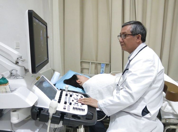
3.1 Steps to perform The patient is performed transthoracic or transesophageal echocardiography like conventional echocardiography. Place a needle into the vein. Acoustic foaming can be self-made acoustic foams or pre-manufactured sound blocking agents. Self-made contrast agents are created from common infusion solutions, this contrast is quite large, so most only reach the right heart chamber very little through the pulmonary capillaries to enter the left heart chamber. Should be used mainly in the evaluation of the right heart, intracardiac shunts ... Pre-manufactured contrast agents: Injected directly into the blood vessels, they are very small in size so they can pass through the pulmonary capillary into the lungs. left ventricular chamber. Therefore, these substances are mainly applied to the left heart: clarifying the chambers of the heart, assessing the movement of the left ventricular walls and myocardial perfusion. Turn on the camera to record a video of the process to review and analyze the image, the direction of the acoustic foam. Observe the course of the air bubbles and evaluate the echocardiographic results. 3.2 Complications after contrast echocardiography Normally, if transthoracic ultrasound, this technique has no complications, only a few cases of people with congenital heart disease have mild headache symptoms, Rest a few minutes will help. In case of transesophageal ultrasound: The throat may be sore for several hours, rarely the ultrasound tube scratches the inner throat. During the ultrasound, you may experience breathing problems due to the sedation or the amount of oxygen you breathe.4, Where is the echocardiogram?
Contrast echocardiography is a technique that looks simple, but it is not, because the heart works continuously, so it is difficult for the survey to accurately diagnose the disease, requiring the doctor to be a good professional. , rich experience, along with advanced equipment system for clear images.Contrast echocardiography helps to accurately diagnose cardiovascular diseases, serving interventions in disease treatment, so now Vinmec Phu Quoc hospital deploys echocardiography technique.
The advantages of choosing Vinmec Phu Quoc hospital:
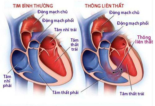
The technique is performed by Doctor Luong Vo Quang Dang - Cardiologist - Head of Department of Medical Examination & Internal Medicine, with years of experience in medical examination and treatment. and cardiology, rich experience in medical examination and treatment for many domestic and foreign patients. Full and advanced medical equipment for medical examination and treatment Very high accurate diagnosis rate Early detection of cardiovascular diseases, especially congenital diseases, and treatment interventions are very important, patients Congenital heart disease for a long time causes complications such as heart failure, high blood pressure... Contrast echocardiography is an effective and safe method to help screen and detect cardiovascular diseases early, especially congenital heart disease.
Please dial HOTLINE for more information or register for an appointment HERE. Download MyVinmec app to make appointments faster and to manage your bookings easily.


