This is an automatically translated article.
The article was professionally consulted by Specialist Doctor I Nguyen Thanh Hai - Doctor of Radiology - Department of Diagnostic Imaging and Nuclear Medicine - Vinmec Times City International General Hospital.The lateral chest radiograph is a technique that helps to add additional information to the straight chest radiograph to diagnose and detect thoracic pathology.
1. What is a lateral chest X-ray?
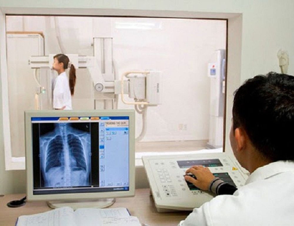
Chest X-ray is a technique that uses X-rays to reconstruct images of the rib cage and the structures inside the chest, including the heart, lungs, and mediastinum. In addition, chest X-ray also helps to image the spine, collarbone, and sternum.Chest radiographs are classified into two main categories, including straight thoracic radiographs and lateral thoracic radiographs. These two techniques are performed in similar steps. The only difference lies in the patient's position relative to the X-ray source. When a lateral chest X-ray is taken, the patient is asked to stand in a slanted position, aligned with the X-ray beam, which is different from that of the radiograph. Straight chest X-ray with the patient standing facing the X-ray beam
Anxillary chest X-ray can help the doctor guide the appropriate treatment and follow-up treatment for the patient through the evaluation of structures such as: :
Heart shadow: size, shape of the heart, and with major vascular arches such as aortic arch, pulmonary artery, superior vena cava, inferior vena cava. In addition, if the patient has pacemakers, artificial valves, defibrillators, and tubes in place, they can also be seen on the lateral chest x-ray because of the contrast. Lungs: The lungs are present on radiographs as bright (black) images due to the presence of gas. In the lateral chest radiograph, the images of tumors, inflammation, fluid retention, and fibrous fibers can be seen, but it is difficult to distinguish which lung lesions are due to overlapping images. Therefore, at this time, it is necessary to have a combination of straight chest radiographs. Skeletal system: the bones that make up the ribcage such as ribs, sternum, and upper spine can be seen on radiographs. Fracture lesions are the most commonly observed group of images on radiographs. SEE ALSO: Why is it necessary to conduct a chest x-ray, straight lung?
Tilt chest X-ray is widely used because it has many advantages such as fast, simple and cheap implementation time. However, lateral thoracic radiographs have some characteristic disadvantages including:
Exposure to X-rays, not suitable for women who are pregnant or suspected of being pregnant. However, the amount of radiation used in a lateral chest X-ray is not very high and usually does not cause serious harm after only a few scans. Small lung lesions may be missed or obscured by the cardiac shadow and rib arches. In some cases, lateral thoracic radiographs do not provide sufficient information for a pathological diagnosis. The clinician will advise the patient to perform a number of other imaging methods, the most common being thoracic computed tomography or echocardiography. Another thing that patients need to remember is that not all abnormalities in the heart and lungs show up on a lateral chest X-ray. The results of chest X-ray film can be different between different medical facilities because of the different imaging methods.

2. Steps to take chest X-ray film
Patients should follow the steps to conduct a lateral chest X-ray to ensure the safety of the patient and the quality of the film. The procedure includes:The clinician appoints an order to take a lateral chest X-ray. The technician receives the order, compares administrative information such as the patient's name, gender, age, diagnosis Adjust the position of the x-ray film, about 1-2 m away from the X-ray source. Enter the imaging room, change clothes and remove jewelry, personal items Ask the patient to stand in a tilted position, aligned with the X-ray beam, bring the chest on the side to be examined close to the machine, arms extended, higher than the head The beam is directed towards the center, towards the lower border of the armpit. Enter the patient's name, gender, and age into the machine. The patient is asked to inhale deeply and hold his/her breath for a few seconds Close the door to the X room. X-ray and start the machine to emit the X-ray beam The patient changes clothes and goes out to the waiting room Print the results and give them back to the patient.
3. When should a chest X-ray film be taken?
When there are symptoms of suspected pathology in the heart, lungs, thorax. Patients are often advised to perform upright and lateral chest radiographs as the first imaging modality. The doctor will advise performing a chest X-ray method when the patient has signs such as:Cough: dry cough, cough with sputum, prolonged cough that does not respond to medication, coughing up blood. Persistent cough and sputum accompanied by unexplained weight loss. Shortness of breath: Acute shortness of breath, appearing on exertion or at rest, signs of exertion such as tachypnea, chest indrawing, rising and falling of the nostrils, retraction of the sternum. High fever: fever accompanied by respiratory symptoms such as cough, difficulty breathing ... Chest pain: pain at any position on the ribcage, pain behind the sternum After thoracic trauma due to falls, falls, traffic accidents.

Currently, chest X-ray is done for almost all patients when hospitalized for treatment, X-ray film provides a lot of useful information for doctors in treatment. However, in order to have accurate and clear diagnostic imaging results, patients need to choose reputable medical facilities that have a system of facilities, modern X-ray machines, and standard weights.
Vinmec International General Hospital has applied the lateral chest X-ray technique in examining and diagnosing the internal structures of the chest including the heart, lungs, and mediastinum. In addition, chest X-ray also helps to image the spine, collarbone, and sternum.
Accordingly, the chest X-ray technique at Vinmec is carried out methodically and in accordance with standard procedures by a team of highly skilled medical professionals, modern machinery system, thus giving accurate results. , contributes significantly to the identification of the disease and the stage of the disease.
Please dial HOTLINE for more information or register for an appointment HERE. Download MyVinmec app to make appointments faster and to manage your bookings easily.


