This is an automatically translated article.
The article was professionally consulted by Specialist Doctor I Tran Cong Trinh - Radiologist - Radiology Department - Vinmec Central Park International General Hospital. The doctor has many years of experience in the field of diagnostic imaging.Two-energy computed tomography (DECT) is a new technique that uses the distinction of matter components based on the difference of two energy levels. Currently, 2-level computed tomography is widely applied in clinical practice and is increasingly proven to have clinical value in helping to diagnose a number of diseases.
1. What is 2-level computed tomography?
Two-energy computed tomography (DECT) is the use of data sets obtained at two energy levels, usually 80kVp and 140kVp, to increase the ability to detect, characterize lesions, and differentiate urinary system stones. , detect tophi nodules, evaluate pulmonary perfusion, exclude bone angiography,...For two-level imaging, the following instruments are available:
Use two lamp heads to emit two at the same time different power levels or use a filter right behind the lamp head. Using one lamp but changing very quickly in two energy levels (GE, Toshiba) Using one lamp but with two receiver layers to create two energy levels Two energy levels of X-ray's main interaction with matter to create diagnostic images mainly thanks to photoelectric effect and Compton scattering. Photoelectric effect is related to the release of energy of outer electrons when entering the innermost or K layer.
Working principle: K-level electron binding energy is also called K-level energy. K-level energy Characteristic for each substance, there is a sudden increase in absorption at this energy level. Most body structures have low energy K levels. From there it is possible to distinguish substances by different K levels at different energy levels between 80kVp and 140kVp.

Chụp cắt lớp vi tính 2 mức năng lượng được ứng dụng rộng rãi trên lâm sàng
Image processing is further analyzed with the 'matter separation' model in the processing software. Specifically breaking down body tissue into substances such as fat, water and iodine. Then detect tissue with a lot of iodine through Iodine map. Virtual drug-free image reconstruction by subtracting the iodine map from the normal CT image.
2. What is a 2-level computed tomography scan for?
Computerized tomography with 2 levels of energy allows detecting and diagnosing a number of diseases such as: Distinguishing urolithiasis, detecting tophi nodules, evaluating pulmonary perfusion,...2.1. Detection of liver damage Iodine mapping in soft tissue organs to study the enhancement of some localized liver lesions such as small tumors, metastases with less vascularity, differentiated from liver cysts, cirrhosis ...
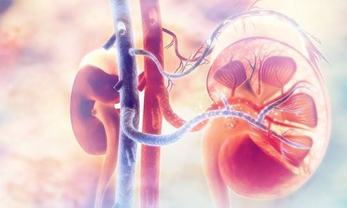
2.2. Renal damage On DECT distinguishes renal cysts with high attenuation on the "no virtual contrast" image but not on the "Iodine map" image. With u, it will be high on the Iodine map because of contrast enhancement
2.3. Distinguishing Types of Kidney Stones The three most common and clinically relevant kidney stones are calcium, uric acid, and struvite. The spectrum of uric acid is weaker than calcium and struvite, so DECT can be distinguished, giving the appropriate treatment for the patient.

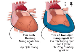
Trường hợp thuyên tắc động mạch phổi, DECT cho thấy hình ảnh tắc mạch máu phổi
2.5. Early detection of gout Assessing gout through urate crystal deposition in joints is an important and meaningful application for early diagnosis of gout. Therefore, the latest Gout diagnostic guidelines have recently included the DECT criteria.
2.6. Detection of bone contusion Bone contusion can be assessed on DECT using the
2.7 bone exclusion technique. Detection of true cerebral hemorrhage DECT is used to distinguish between true hemorrhage and hyperattenuating images due to stagnation of iodinated contrast after revascularization intervention in acute stroke. In actual bleeding there is no enhancement on the iodine map. Thus, it helps in the diagnosis of true cerebral hemorrhage.
Two-energy computed tomography is the current method, the procedure is simple, it helps in early diagnosis of some diseases of great clinical significance.
Vinmec International General Hospital with a system of modern facilities, medical equipment and a team of experts and doctors with many years of experience in medical examination and treatment, patients can rest assured to visit. examination and treatment at the Hospital.
Please dial HOTLINE for more information or register for an appointment HERE. Download MyVinmec app to make appointments faster and to manage your bookings easily.
SEE MORE
128-slice CT scanner at Vinmec Relation between X-ray and Pregnancy Notes on coronary angiography