This is an automatically translated article.
The article is professionally consulted by Master, Doctor Nguyen Van Huong - Department of Diagnostic Imaging - Vinmec Danang International General Hospital.
Testicular ultrasound is a common medical technique but plays an important role in the examination, diagnosis and treatment of diseases related to the testicles - an important part of the male reproductive organs. The correct diagnosis and detection of the disease condition has a great impact on the later treatment process.
1. Testicle Overview
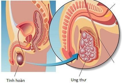
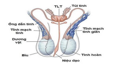
The testicles are a part of the male sex organ whose main function is to produce sperm and secrete the male sex hormone Testosterone that forms male characteristics. Each male has 2 testicles: left and right, located in the scrotum, each weighing about 8-12 grams, 4-5cm long. When noticing abnormal signs in the testicles, doctors will assign the patient to perform testicular ultrasound to have a more specific diagnosis. Testicular ultrasound is a common technique that uses ultrasound waves to capture images of the inside of a male testicle. Combined with blood and semen tests, it will help diagnose pathologies in the genital organs such as epididymitis, varicocele, testicular torsion, hydrocele, etc.
2. Testicular ultrasound for what?
As mentioned above, testicular ultrasound helps doctors:Check for a lump or painful swelling in the testicle.
● Find or check the site of infection in the testicles
● Evaluate for spermatic cord torsion (a condition that causes blood supply to the testicles to be affected).
● Monitor whether testicular cancer has recurred or not.
Diagnosis of undescended testicle
● Check for fluid/blood in the scrotum, fluid in the epididymis or pus in the scrotum.
Check for trauma to the genitals (especially testicles)

Trắc nghiệm: Bạn biết bao nhiêu về tinh dịch?
Tinh dịch là một phần quan trọng quyết định đến khả năng sinh lý, sinh sản của nam giới. Liệu bạn đã hiểu được bao nhiêu về thành phần quan trọng này? Hãy cùng thử tài thông qua các câu hỏi nhanh về tinh dịch sau đây nhé!
Bài dịch từ: webmd.com
3. Testicular ultrasound helps detect which diseases?
Although testicular ultrasound is a fairly simple diagnostic method, it plays a very important role in the detection and treatment of diseases related to the male genital organs, namely some pathologies. The following:Varicose veins:
Varicocele (also known as “varicose veins of the testicle/scrotal varicocele”) is abnormally dilated veins above the testicles causing swelling. Heaviness in the scrotum and reduced sperm count, which can lead to infertility. An ultrasound examination of the testicles will show the dilated tufts of the spermatic cord, causing blood to stagnate and not circulate.
● Testicular torsion
Testicular torsion is a phenomenon in which the testicle is twisted around its axis, causing blockage of blood vessels, causing testicular swelling, atrophy or congestion, and can lead to testicular necrosis. The disease can occur at any age but is most common in men 10-25 years old. The testicular ultrasound will help the doctor assess the severity of testicular torsion to have a timely treatment plan.
● Orchitis
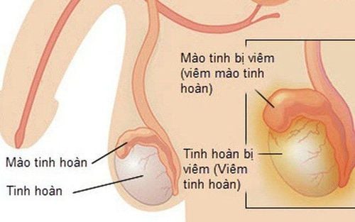
Orchitis is an inflammation of one or both testicles due to the penetration of bacteria, viruses, staphylococcus aureus, bacilli. Or it can also be due to trauma, acute infection after epididymitis. The typical symptoms of orchitis are painful swelling of the testicles, heaviness in the scrotum, pain when urinating or blood in urine, etc. When an ultrasound scan of the testicles will clearly see the foci of inflammation, swelling, Membrane effusion.

Epididymitis is painful swelling and inflammation in the testicles, specifically the coiled tube at the back of the testicle. It is caused by a retrograde infection from the bladder and prostate, or by a sexually transmitted infection. Ultrasound images will show inflammation in the epididymis.
● Epididymal cyst
Epididymal cyst is a blockage of the vas deferens in the epididymis, forming a benign watery mass that appears at the epididymis. This mass can grow over time and can make a man infertile.
● Testicular injury
Is a fairly common condition when men exercise with intensity and accidentally hit the genitals, causing injury. In this case, ultrasound imaging of the testicles can help doctors detect and assess the extent of injury to this vital organ.

4. Normal testicular ultrasound technique
Testicular ultrasound is an assistive technique to help observe and evaluate the pathological condition inside the testicles. It can also show the vas deferens that connect the testes to the prostate. The transducer sends sound waves to a computer that plays back images of the organ being scanned.● Preparing for ultrasound
To perform an ultrasound, the patient hardly has to prepare anything. If a biopsy or other test is performed at the same time during the ultrasound, the patient may need to sign a consent form.
● Conduct ultrasound
Step 1: Before the procedure begins, the patient will be asked to take off his pants from the waist down and put on a temporary gown.
Step 2: The patient will be instructed to lie supine on the ultrasound table and use a folded towel to cover the penis and raise the scrotum.
Step 3: A gel will be spread by the technician on the scrotum to reduce friction on the skin and help the transducer get a better image.
Step 4: The patient needs to lie still while the probe is pressed against the skin in the testicle and moved through the scrotum several times. If the test is used to assess the extent of trauma or to find out the cause of testicular pain, you may feel pressure as the transducer gently presses against the affected area. If a biopsy is needed during the ultrasound, you may feel some discomfort while taking the sample.
Step 5: At the end of the procedure, the technician wipes the gel off the patient's skin and helps the patient sit up.

5. Testicular ultrasound evaluation results
Normal testicle image: When the testicle is in normal shape and size, it will be <5cm high, about 3cm wide, about 2cm thick. In addition, a normal testicle on ultrasound will:● No sign of benign or malignant tumor
● No sign of infection or swelling, epididymitis
● No torsion of the spermatic cord, sperm blood supply is normal
● There is no sign of fluid, blood, or pus in the scrotum.
Testicles are evaluated for abnormality when:
● Identify a tumor in the testicle or signs of testicular cancer.
● There are signs of infection or swelling, epididymitis.
● Seeing that the spermatic cord is twisted, the testicle is not supplied with blood, causing atrophy, the risk of necrosis.
● No testicle or only one testicle in the scrotum.
● See fluid, blood, pus in the scrotum or see fluid in the epididymis.
● See hernia in the scrotum.

Master Doctor Nguyen Van Huong used to be a Lecturer in Medical Imaging at Danang University of Medicine and Pharmacy, many years of experience working in hospitals such as Cancer Hospital; Da Nang Hospital; Da Nang Obstetrics and Gynecology Hospital.
With his dedication to his work, dedication to the medical profession in general and the specialty of diagnostic imaging in particular, Dr. Huong is always loved and trusted by patients.
To register for examination and treatment at Vinmec International General Hospital, you can contact Vinmec Health System nationwide, or register online HERE.
Recommended video:
Infertility treatments: Hope for infertile and infertile couples
MORE
10 reasons for alarming testicle pain Signs of undescended testicles in children Epididymitis: Causes, symptoms, diagnosis and treatment


