This is an automatically translated article.
To detect lung lesions, in addition to chest x-ray and computed tomography, patients are also assigned to aspirate pleural fluid and biopsies to get more diagnostic information.
1. Aspiration of pleural fluid
1.1. When is pleural aspiration indicated?
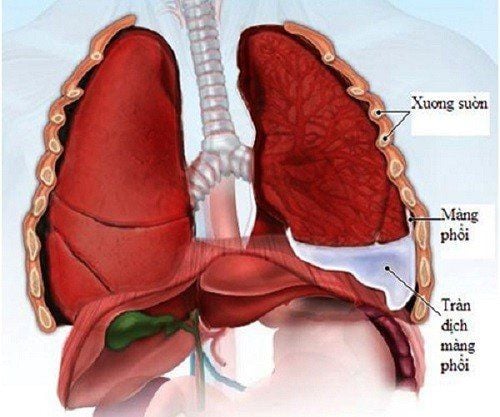
Pleural effusion is a technique that uses a needle to aspirate fluid when a patient has pleural effusion. This procedure is performed to diagnose or treat disease in the following cases:
Diagnosis: Indicated for most patients with new symptoms of pleural effusion or when the cause is uncertain. . Treatment: Indicated to relieve the symptoms of the disease.
1.2. The technique of aspiration pleural fluid
Procedures for performing pleural effusion technique are as follows:
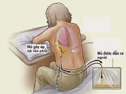
Step 1: Confirm the degree of pleural effusion by tapping on the chest wall and observing X-ray, CT, and ultrasound images to determine the location of the pleural effusion. position of translation. Step 2: Needle puncture in the midline of the scapula, superior edge of the rib at the superior space of the effusion. Then, mark the puncture point and disinfect the skin. Step 3: Using a 25-gauge needle, inject local anesthetic. Switch to a larger (20 or 22-gauge) needle. At the same time, inject anesthetic slowly until it reaches the parietal pleura, this area needs to be injected with anesthetic the most, because this is a very sensitive location. Step 4: Continue to push the needle until the pleural fluid is aspirated, then record the depth of the needle.

Chọc hút dịch màng phổi là kỹ thuật dùng kim để hút dịch khi bệnh nhân bị tràn dịch màng phổi.
1.3. Risks of aspiration of pleural fluid
In the process of performing pleural aspiration technique, there may be unintended risk complications such as:
Pneumothorax: This is the most common risk of pleural aspiration; Bleeding: The patient coughed up blood from the puncture of the lung; Edema due to enlarged lungs or low blood pressure; Hemothorax due to damage to the intercostal blood vessels: It is caused by the incorrect placement of the needle, touching the blood vessels and intercostal nerves; Poke the spleen or liver; Improper anesthesia of the parietal pleura; The patient fainted due to the vagal reflex.
2. Pleural biopsy
2.1. What are the indications for a pleural biopsy?
This is a technique that uses a pleural biopsy needle to collect a patient sample under the guidance of a CT scan. The obtained fluid will provide more information in the diagnosis of the disease.
The advantage of this technique is that it is less invasive and can diagnose benign or malignant tumors. In particular, the recovery time is fast, the patient can soon return to daily activities.
2.2. Procedure for performing a pleural biopsy
The procedure for performing a needle biopsy of the pleura under the guidance of the CT scanner is as follows:
Step 1: CT scan to locate the mass. Then, mark and disinfect the suspected lesions. Step 2: Anesthetize the injection area. Step 3: Make a very small incision in the skin at the pre-marked position, insert the biopsy needle to aspirate the specimen through the guidance of the CT technique. Step 4: Withdraw the biopsy needle after taking the required amount of specimen. Step 5: When the procedure is complete, the area where the biopsy needle was inserted will be gently pressed (to help stop bleeding) and bandaged.

Sinh thiết màng phổi bằng kim dưới hướng dẫn của CT scanner.
2.3. Risks when performing pleural biopsy technique
Performing a pleural biopsy may pose some risks such as:
Risk of infection: Needing antibiotic treatment; Bleeding at the biopsy needle insertion site when the patient coughs: This is rare; Presence of gas leak at the biopsy site: This complication can cause pneumothorax and lead to atelectasis. Patients present with symptoms of atelectasis: Shortness of breath, shortness of breath, rapid pulse, sharp pain in the chest or shoulder when breathing. Most pneumothorax will resolve on their own. If the condition does not improve, the doctor will use a small catheter placed in the chest cavity to guide the air out. In summary, aspiration and pleural biopsy are "golden" medical techniques that help doctors diagnose and treat lung diseases. However, these techniques can bring certain risks, so patients need to choose reputable medical facilities, leading in the field of imaging to perform the examination. Currently, Vinmec International General Hospital is one of the leading prestigious hospitals in the country, trusted by a large number of patients for medical examination and treatment. Not only the physical system, modern equipment: 6 ultrasound rooms, 4 DR X-ray rooms (1 full-axis machine, 1 light machine, 1 general machine and 1 mammography machine) , 2 DR portable X-ray machines, 2 multi-row CT scanner rooms (1 128 rows and 1 16 arrays), 2 Magnetic resonance imaging rooms (1 3 Tesla and 1 1.5 Tesla), 1 room for 2 levels of interventional angiography and 1 room to measure bone mineral density.... Vinmec is also the place to gather a team of experienced doctors and nurses who will greatly assist in diagnosis and detection. early signs of abnormality in the patient's body. In particular, with a space designed according to 5-star hotel standards, Vinmec ensures to bring the patient the most comfort, friendliness and peace of mind.
Please dial HOTLINE for more information or register for an appointment HERE. Download MyVinmec app to make appointments faster and to manage your bookings easily.


