This is an automatically translated article.
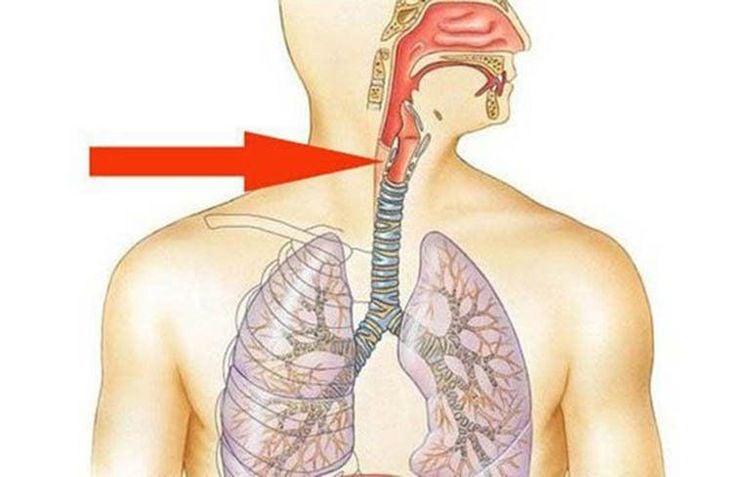
Esophageal webs are indentations of the esophageal wall that can partially narrow the lumen of the esophagus. Initially, the esophageal web is usually asymptomatic, but occasionally patients may experience intermittent dysphagia with solid foods.
1. Web esophagus is what disease?
Esophageal web or esophageal septum is a thin structure that partially narrows the lumen of the esophagus. Patients with esophageal septum are usually asymptomatic. However, others have intermittent dysphagia with solid foods rather than liquid foods.
The esophageal septum is a thin membrane (<2 mm) eccentrically protruding into the lumen of the esophagus. Histologically, the esophageal septum is covered with squamous epithelium and most commonly occurs anteriorly of the esophagus, causing localized esophageal stricture in the posterior region.
The true incidence of esophageal septum is difficult to determine in practice, as most patients are asymptomatic. However, an observation of esophageal septum has been reported in 5 to 15% of patients undergoing barium esophagography for dysphagia.
2. What causes the esophageal diaphragm?
The pathogenesis of the esophageal septum is so far unknown. It is hypothesized that the esophageal septum has a secondary origin after chronic damage from gastroesophageal reflux. However, a congenital or primary origin of the advanced esophageal septum has also been suggested.
Evidence that exposure to esophageal acid causes esophageal septa and esophageal rings is supported by studies showing a reduced risk of recurrence of the condition if patients are treated with inhibitors. acid. At the same time, a radiographic study demonstrated that the progression of the esophageal septum causing progressive narrowing of the esophagus was due to esophageal reflux.

Web thực quản là một cấu trúc mỏng làm hẹp lòng thực quản một phần
3. What are the signs and symptoms of esophageal septum?
Most patients with esophageal septum are asymptomatic. In contrast, symptomatic patients often present with difficulty swallowing solid foods, especially evident with hard foods. Dysphagia is often intermittent and patients will occasionally need to change the way they eat (eg, chew food more thoroughly).
In addition, the manifestations and severity of symptoms with esophageal septum also depend on the inner diameter of the esophageal lumen. Patients with esophageal strictures smaller than 13 mm will often have difficulty swallowing solid foods.
Occasionally, patients present with acute-onset dysphagia or complete inability to swallow saliva due to food contamination. In rare cases, patients with esophageal septum have been associated with Plummer-Vinson syndrome with associated clinical features of iron deficiency anemia.
4. How to diagnose esophageal diaphragm
The esophageal septum causing narrowing of the esophagus is directed when the patient has a long history of dysphagia with solid foods. However, for definitive identification, the patient should be indicated for diagnosis by barium swallow and/or upper gastrointestinal endoscopy to evaluate dysphagia or rule out other upper gastrointestinal symptoms.
Barium esophagogram On barium esophagogram, the esophageal Web appears as a thin border (<3.0 mm) in circumference several centimeters across the hiatus of the diaphragm. The muscle rings are smooth, symmetrical narrow slits 3 to 5 mm axially with a variable aperture during fluoroscopy examination.
However, if the esophagus is not fully dilated, the esophageal septum or esophageal ring, if present, causes a discreet narrowing of the lumen that can easily be missed.
Upper Gastrointestinal Endoscopy With endoscopy, the esophageal septum appears as a thin, smooth membrane that usually does not obstruct the bronchoscope.
Upper gastrointestinal endoscopy is less sensitive than barium esophagography for detecting esophageal rings and diaphragms. In particular, the esophageal septum is often missed during upper gastrointestinal endoscopy because they are located near the upper esophageal sphincter.

Web thực quản thường không có triệu chứng nhưng đôi khi có thể xuất hiện với chứng khó nuốt
5. Treatments for the esophageal diaphragm
The esophageal septum is often easily ruptured during diagnostic endoscopy when the interventional endoscope crosses the membrane. However, in patients who already have narrowing of the esophagus, the patient needs to be dilated to restore normal gastrointestinal function.
After esophageal dilation, the patient should be on maintenance therapy with a standard once-daily dose of a proton pump inhibitor for 6 weeks. For patients with esophageal septum and evidence of gastroesophageal reflux (eg, patients with frequent heartburn, or erosive esophagitis on endoscopy), this therapy should be continued indefinitely. , which effectively relieves dysphagia and prevents recurrence of symptoms.
In addition, if the patient does not respond to conventional esophagogastric dilation, or has recurrent choking or dysphagia, the patient may need to be indicated to remove the esophageal septum by more specialized methods, such as esophagectomy. Endoscopic incision therapy uses electrocautery, endoscopic laser cutting, or biopsy forceps.
In summary, webs of the esophagus are usually asymptomatic but can sometimes present with intermittent dysphagia with solid foods if significant narrowing of the esophageal lumen is present. At this point, barium film will be especially helpful in determining the cause of the esophageal lumen obstruction. In addition, esophagogastroduodenoscopy is also necessary to diagnose as well as decide to intervene locally, cut the esophageal septum and effectively widen the esophageal lumen for the patient.
Please dial HOTLINE for more information or register for an appointment HERE. Download MyVinmec app to make appointments faster and to manage your bookings easily.
References: cedars-sinai.org, sciencedirect.com, msdmanuals.com, emedicine.medscape.com


