This is an automatically translated article.
Posted by Master, Doctor Pham Manh Chung - Head of Imaging Department - Department of Diagnostic Imaging - Vinmec Ha Long International Hospital
Magnetic resonance imaging of nerve fiber bundles, also known as tension diffusion magnetic resonance imaging. This is a modern imaging technique, applied to detect neurological diseases in which nerve axons are damaged.
1. Purpose of Nerve Fiber Bundle Magnetic Resonance Imaging
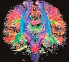
Difusion tensor imaging (DTI) or magnetic resonance imaging (Tractography) is a modern imaging technique in magnetic resonance imaging to evaluate pathology. nerve. DTI is an MRI technique that uses anisotropic diffusion to estimate the white matter axonal organization of the brain. Fiber bundle magnetic resonance imaging is a 3D reconstruction technique for the evaluation of nerves using data collected by tension imaging.
The purpose of this pulse scan is to evaluate axonal lesions of the brain, spinal cord or to find a relationship between the lesion and the nerve fiber bundles to avoid damage to the nerve fiber bundles when treating the lesions .
Applications of neurofibrillation are thriving because the technique is very sensitive to changes at the cellular and microstructural levels.
2. When should magnetic resonance imaging of nerve fiber bundles be performed?
Indication of nerve fiber bundle magnetic resonance imaging is indicated when the specialist suspects that the lesion has compression, invasion into the nerve fiber bundles or the doctor wants to evaluate the relationship of the lesion to other nerve fibers. fiber bundles for an appropriate treatment plan to avoid damage to the motor or sensory bundles.
Specifically indicated in the following cases:
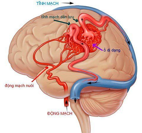
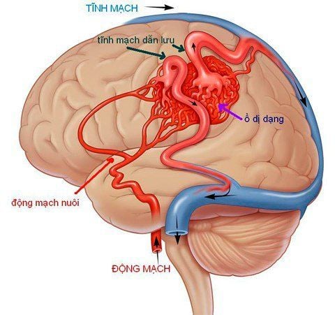
Brain tumors that invade or lie next to axon bundles Multifocal sclerosis, white matter lesions in infarction, bleeding,... Allergic lesions cerebrovascular pattern, it is necessary to find the relationship of the malformation with the axon bundle. Congenital malformations: ectopic gray matter, cleft encephalopathy.. Epilepsy Cerebral palsy. cochlear poles, automatic intramuscular pumps (absolute contraindication) In people with ferromagnetic metals (relative contraindications) People with fear of light, fear of lying alone Inability to lie still (yes) sedatives may be used).
3. Magnetic resonance imaging procedure of nerve fiber bundles
Step 1: Prepare the patient
The patient is checked for magnetic resonance safety before being brought into the imaging room Lie on his back on the table Select and position the receiver Move the table into the machine compartment and locate the area Taking the step 2: Performing the technique
Location capture:
Select the diagnostic pulse sequences suitable for examination purposes, usually T1, T2, FLAIR. The cutting direction includes horizontal (horizontal), horizontal (horizontal) and vertical (vertical) cutting. Perform a conventional diffusion pulse sequence and calculate the apparent diffusion coefficient map (ADC map) to assess the lesion. In addition, each detected lesion can perform the necessary pulse sequences, eg T2* sequences, to find out if the lesion is bleeding.
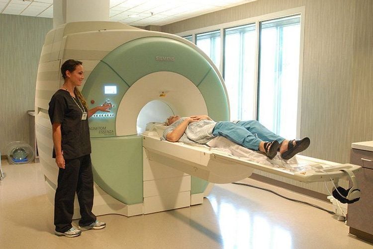
Select the tensor pulse sequence and select the direction depending on the direction of the axon to be examined. Run each pulse and process the images obtained on the workstation screen, select the necessary images to reveal the pathology for film printing. The doctor analyzes the results and makes a diagnosis. Step 3. Monitoring
During the procedure: monitor pulse, blood pressure, consciousness through the camera and/or electrocardiogram on the screen After the procedure: let the patient wait 30 minutes in the waiting room to follow up. Thanks to the development of modern medicine, today, lesions and diseases related to nerve fibers and nerves are detected and diagnosed thanks to the technique of magnetic resonance imaging of nerve fiber bundles. However, in order to provide the best image for a suitable treatment regimen, patients should choose addresses with quality modern magnetic resonance imaging machines.
To meet the needs of examination, imaging and treatment of diseases related to the nervous system as well as many other diseases, Vinmec International General Hospital has brought modern equipment such as: magnetic resonance imaging (MRI), computed tomography (CT) scan, X-ray, .. in medical examination and treatment, diagnostic imaging, including neurological-related diseases. Therefore, the diagnostic imaging results bring true and clear images for effective medical examination and treatment.
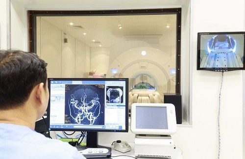
Especially, before taking magnetic resonance imaging, customers will be evaluated by leading medical experts about their medical history, personal and family history and magnetic resonance safety check (check list MRI). ) to ensure the safety of the client before entering the magnetic resonance room, to limit complications after the scan.
Please dial HOTLINE for more information or register for an appointment HERE. Download MyVinmec app to make appointments faster and to manage your bookings easily.


