This is an automatically translated article.
The article was written by Doctor of Radiology, Department of Diagnostic Imaging - Vinmec Central Park International General Hospital.IntraVascular UltraSound is internationally known as IntraVascular UltraSound (IVUS for short). This is one of the modern and advanced diagnostic imaging methods, which is a valuable invention for the medical field, helping doctors evaluate and observe the structure of blood vessels, thereby detecting signs and symptoms. abnormalities and prompt treatment.
1. What is Intravascular Ultrasound (IVUS)?
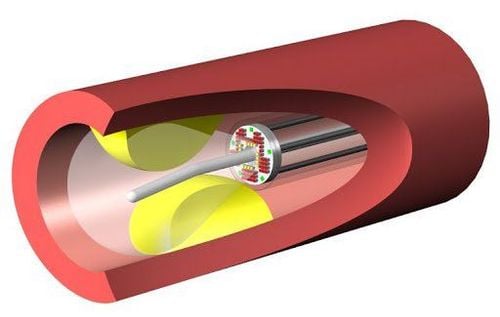
Intravascular ultrasound is an ultrasound technique that uses a special catheter to observe and evaluate the structure of blood vessels. One end of the catheter is attached to a transducer that emits ultrasound waves, and the other end is connected to an image acquisition and processing system. The catheter will be threaded through a wire straight into the lumen of the blood vessel, to the location to be surveyed and recorded blood vessel images. Through the obtained images, doctors will assess the condition of blood vessels and diagnose related diseases.
2. Value of intravascular ultrasound
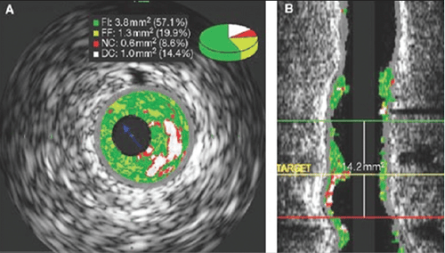
Intravascular ultrasound is an imaging method applied in cases of suspected vascular disease, especially coronary artery disease. The results obtained from intravascular ultrasound reflect in detail, specifically the condition of blood vessels, including: size, shape, atherosclerotic lesions, calcifications, thrombosis... In cases where it is difficult to identify coronary artery disease, in addition to conventional coronary angiography, doctors often assign intravascular ultrasound to have accurate diagnosis results.
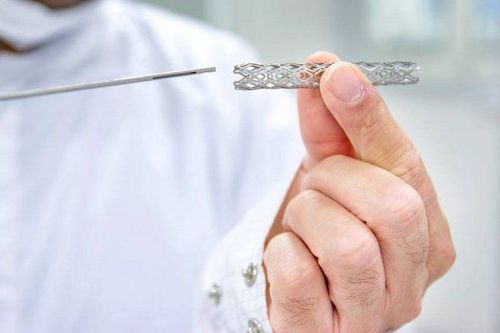
Through intravascular ultrasound, the doctor can give appropriate treatment such as: stent placement, stent size, necessary tools for the intervention process...
After finishing During treatment, intravascular ultrasound is also indicated to evaluate the effectiveness of treatment, investigate the cause of restenosis in cases of restenosis after stent intervention.
In summary, intravascular ultrasound plays a very important role in the diagnosis, treatment, and recovery after treatment of patients with diseases related to blood vessels.

Siêu âm trong lòng mạch có vai trò quan trọng trong chẩn đoán và điều trị bệnh lý mạch máu
3. Notes before intravascular ultrasound
Before having an intravascular ultrasound, the patient should inform the doctor about:
Current medications Allergic conditions Recent medical conditions Chronic conditions The patient needs to remove jewelry. , metal objects, including dentures so as not to interfere with the procedure.
Intravascular ultrasound is often combined with coronary angiography under the light in the X-ray room, so women who suspect that they are pregnant need to inform their doctor before performing the procedure.
4. Intravascular ultrasound procedure
The IVUS catheter is a thin tube. One end of the tube attaches to the ultrasound transducer, the other end connects to a computer to convert the signal from the transducer into an image on the screen. Intravascular ultrasound, like other ultrasound techniques, also uses high-frequency sound waves to image the structure of blood vessels.
The patient will be placed on the procedure table, attached devices to monitor blood pressure, heart rate, blood oxygen to conduct combined coronary angiography.
The technician will place an intravenous line to give fluids and other medications if needed. Many patients require a moderate dose of sedation.

Một đầu của ống gắn vào đầu dò siêu âm trong lòng mạch
Depending on the specific situation, some patients may be assigned general anesthesia, while the rest will be given local anesthesia. The doctor will disinfect the catheter site and cover it with a surgical cloth. A guide is inserted into the forearm or femoral artery. The catheter is threaded into the guide device, going to the site of interest on a flexible wire. This process is carried out under the guidance of the dimming curtain. Here, ultrasound waves will create an image of the blood vessel structure, reflected back to the display screen.
After getting the desired image, the doctor will proceed to remove the catheter and guiding device from the patient's body. The insertion site will be pressed to stop bleeding.
5. Notes after intravascular ultrasound
After removing the catheter and applying pressure, applying pressure to stop bleeding, the patient needs to lie down with the leg straight for at least 24 hours. If your doctor uses a device that seals the catheterization site in your thigh, you may be able to get up and walk around sooner. Side effects of sedatives, anesthetics, so patients will feel sleepy, sleep a lot. The patient is checked for blood pressure, heart rate, and bleeding status within a few hours after the ultrasound. Depending on the specific technique, the patient will be required to stay in the hospital for monitoring for 24-48 hours After going home Patients should rest, avoid exercise, strenuous exercise for at least 24 hours. Should drink a lot of water The patient may feel pain, bruised skin at the catheter insertion site If there are any abnormal signs, bleeding ... should immediately notify the doctor.

Một số trường hợp, bệnh nhân sẽ được yêu cầu ở lại bệnh viện theo dõi
Vinmec International General Hospital is a high-quality medical facility in Vietnam with a team of highly qualified medical professionals, well-trained, domestic and foreign, and experienced.
A system of modern and advanced medical equipment, possessing many of the best machines in the world, helping to detect many difficult and dangerous diseases in a short time, supporting the diagnosis and treatment of doctors the most effective. The hospital space is designed according to 5-star hotel standards, giving patients comfort, friendliness and peace of mind.
Please dial HOTLINE for more information or register for an appointment HERE. Download MyVinmec app to make appointments faster and to manage your bookings easily.


