This is an automatically translated article.
The article is professionally consulted by Master, Doctor Vu Thi Hau - Radiologist - Department of Diagnostic Imaging and Nuclear Medicine - Vinmec Times City International General Hospital.Computed tomography (CT) scan of the ear - sclera is an imaging method of high value in the subclinical examination of diseases related to the ear - sclera, especially of great value in assessing Inner ear structures before cochlear implantation in children for children with congenital deafness, sudden deafness, or evaluation of the skeletal structures of the ear. This method can evaluate the morphology, anatomy as well as pathologies related to the components of the ear such as the outer ear, middle ear and inner ear, detect common diseases such as otitis media, cholestetoma , otitis externa in children, ... as well as bone diseases such as fibrous dysplasia, osteoma, calcification of the cochlea, vestibular and semicircular canals, can interfere with the insertion process. electrode.
For the following cases of cochlear implantation, it is often not possible to evaluate with an MRI because the magnetic force of the machine can affect the electrode. Therefore, routine CT and X-ray are good methods to evaluate the position of the post-implantation electrode. X-rays are often used, especially in children, because of their short duration and low radiation dose. However, to accurately assess the position of the electrode, the X-ray film is still limited compared to the CT scan. For adult cases or suspected electrode deviation that cannot be assessed by X-ray, CT is the first choice.
1. Indications and contraindications for ear-bone computed tomography
1.1. Point
Congenital abnormalities of the ear morphology or congenital deafness. Injury, inflammation, ear infection. Hearing loss, dizziness, tinnitus. Ear damage caused by u. Check again after placing the cochlear electrode.1.2. Contraindications
The method of computed tomography of the ear - bone stone has no absolute contraindications but only relative contraindications due to the low risk of radiation contamination. If the patient is unable to perform the technique, an MRI can be substituted.Pregnant women under 3 months need to consider before taking the scan, if done, it is necessary to take measures to protect the abdomen. Small children should be limited, should only be taken in case of necessity.
Nhiễm trùng tai được chỉ định chụp cắt lớp vi tính tai - xương đá
2. What should the patient do to prepare before an ear-bone computed tomography scan?
Prepare a full medical record, order paper for computed tomography of the ear - stone bones. Exploiting some basic clinical information related to medical examination and treatment. Psychological preparation before implementing the method should not be too worried to affect psychology. During the shooting process, you need to lie still so that the captured image does not cause noise. Remove metal objects that interfere with the tomography image such as hairpins, earrings, necklaces. It is recommended to fast for 4 hours before the scan for cases requiring injection. If the assessment is taken before or after electrode placement, fasting is not necessary.3. Medical supplies in preparation for ear-bone computed tomography scan
Computerized tomography machine with appropriate range (16 ranges or more) Dedicated electric pump to control injection velocity. Image storage system. Needle capacity 10ml, 20ml. Injection type 18-20G (if injection is required). Cases requiring injections are decided by the radiologist and discussed with the clinician. Water soluble iodine contrast agent. Antiseptic solution for skin and mucous membranes. Distilled water or physiological salt. Gloves, hat, mask, surgical gauze Set of bean trays, surgical forceps. First aid kit in case of complications after contrast injection.
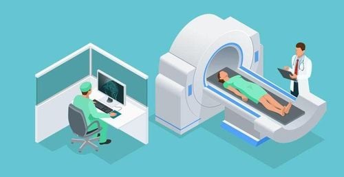
Chụp cắt lớp vi tính tai - xương đá
4. Steps to conduct computed tomography of ear - bones
First, the patient is explained the imaging process, no need to fast if the scan does not inject contrast.The patient enters the imaging room, puts the patient in the supine position on the scanning table, the head inside the skull support is facing the gantry, the legs are facing out.
Enter the patient's information, select the appropriate procedure for the ear - stony bones and appropriate parameters.
The scanning process for modern multi-segment CT scanners in the stony ear region is completed within 1-2 minutes. Slices are visualized in axial and coronal orientations for anatomical evaluation and pathologic evaluation.
Shooting technique
Select the axial cross-section:
The cutting plane should be parallel to the hard palate. Take from the styloid to the superior border of the temporal bone. The lower part from the lower border of the base of the skull, the upper part ends at the upper edge of the auricle or the end of the mastoid bone. Section thickness less than 1mm. Transverse view:
Change the patient's lying position to maximal supine or maximal supine position. Capturing the position of the cutting plane perpendicular to the horizontal plane, the position from the anterior margin to the posterior border of the stony bone. The thickness of each cutting layer should not be more than 1mm. Spiral cutting is recommended, the jump is equal to the thickness of the spiral. Previously, for 2-row CT scanners, it was necessary to cut in two directions, now with the development of technology, multi-row CT scanners have been born, contributing to reducing the time of shooting, reducing the level of radiation contamination as well as the radiation exposure. like providing the best image quality.
Print film
Assess the results of the scan
Before placing the electrode: Assess the structure of the ear is normal or abnormal, there are changes that affect the surgical process or not. After placing the electrode: how is the position of the electrode placed? Through imaging, describe the location, structure, size, and spread of the lesion. Compare the results of computed tomography with clinical examination. Provide diagnostic guidelines and other coordinated examination methods if necessary.
5. Complications and treatment
When taking computerized tomography of the ear - bone stone for children, there are quite a lot of difficulties because the baby refuses to cooperate during the imaging process. Depending on the case, it is possible to use sleeping pills or psychotropic drugs in an appropriate dose. In case the patient is not supine for the transverse CT scan, the image can be reconstructed from the cross-sectional direction. To facilitate reconstruction, the thinnest possible cross-section should be taken. Patients with allergy or contrast shock, depending on the severity, have different management (only for the case of intravenous contrast injection).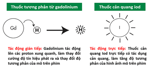
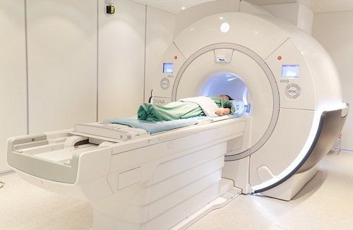
Chụp cắt lớp vi tính tai - xương đá có tiêm thuốc đối quang từ
6. Why should computed tomography (CT) ear - stone bones at Vinmec
Vinmec International General Hospital owns the most modern equipment system for diagnosis and treatment in Vietnam today. The machine systems used in the imaging department are imported from abroad, the quality is equivalent to that of leading hospitals in developed countries such as the UK, USA, Japan,... The goods will be shortened but the picture quality will be the best. Not only that, when performing computed tomography of the ear - bones at Vinmec, you will be examined by a team of experienced and highly qualified medical doctors.During its operation, Vinmec always promotes the training, scientific research and skill improvement of young doctors. In addition, the medical system at Vinmec is managed and operated according to international standards, with a civilized and polite medical examination and treatment space, ensuring maximum sterilization. Vinmec is committed to bringing the best medical examination and treatment services to customers.
Master. Doctor. Vu Thi Hau has many years of experience in the field of diagnostic imaging including magnetic resonance, CT and ultrasound. Graduated from Hanoi Medical University in 2014 with a BSNT and trained in France for 1 year. Currently engaged in the field of diagnosis of neurological, head and neck, ear - bone, musculoskeletal, digestive and urological diseases.
Contact Vinmec here for advice and answers to questions related to computerized tomography (CT) of the ear - bones.