This is an automatically translated article.
The article is professionally consulted by Master, Resident Doctor Nguyen Van Anh - Radiologist - Department of Diagnostic Imaging and Nuclear Medicine - Vinmec Times City International General Hospital.Ultrasound is a basic, non-invasive imaging method that brings a lot of valuable information for diagnosis, monitoring as well as treatment orientation.
1. Ultrasound machine operation
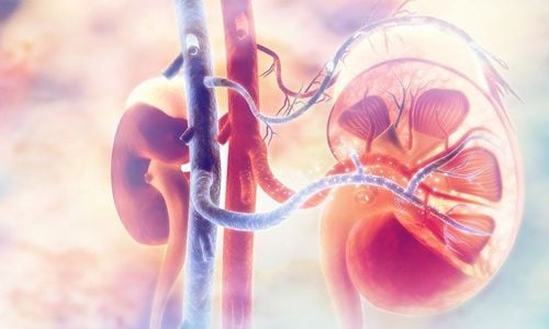
The transducer is the most important part of the ultrasound machine, which is a compact but contains structures capable of transmitting and receiving sound wave signals. When the ginseng wave is emitted, transmitted to the body, to the organ to be examined, the sound wave is partially absorbed, the rest will bounce back to the transducer. Depending on the characteristics of each organ structure, the speed of sound propagation varies. Based on the intensity and time of the reflected sound waves, the computer will analyze and produce the corresponding image.2. Ultrasound image
The outputs of an ultrasound process are images obtained on a monitor, also known as ultrasound images. These images will be transferred to help the treating doctors make the most accurate and comprehensive diagnosis of the damage of tissues and organs, thereby making an appropriate treatment plan.
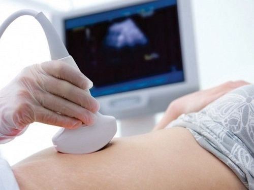
Siêu âm giúp theo dõi được hình ảnh thai nhi
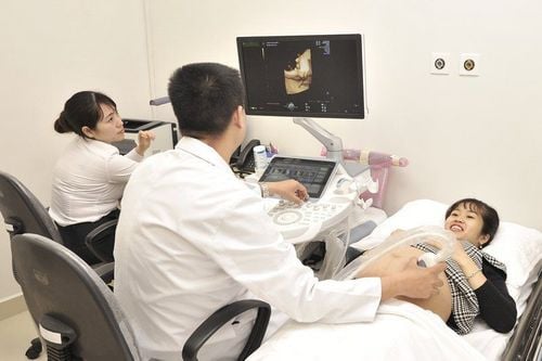
3. Types of ultrasound
● 2D ultrasound: is the most common and basic ultrasound method, helping to provide information about the morphology and structure of organs in the body...● Doppler ultrasound: Is a type of solid ultrasound In particular, they create an image of the blood flowing in the blood vessels, thereby determining the flow, speed and pressure of the blood against the vessel walls.

Các loại siêu âm cho phép chẩn đoán hình ảnh rõ ràng và không xâm lấn
● 4D ultrasound: Similar to 3D ultrasound but the image changes over time, in the form of video, with movement in three-dimensional space. This method is widely applied in fetal ultrasound and gives results superior to other forms of ultrasound. Parents can not only see the shape of the fetus, but also observe the baby's hands, feet, mouth and movements in the womb.
4. Benefits of Ultrasound
● Non-invasive, safe, effective, no side effects.● The patient is not exposed to ionizing radiation as in x-rays or tomography.
● Ultrasound helps doctors see images of soft tissues, which X-rays in other methods do not or cannot show.
● Ultrasound can be performed easily, quickly and at a lower cost than other imaging methods.

Hình ảnh máy siêu âm vú 3D tại Bệnh viện Đa khoa Quốc tế Vinmec
Customers can directly go to Vinmec Health system nationwide to visit or contact the hotline here for support.
Source: webmd.com