This is an automatically translated article.
The article was professionally consulted by Master, Resident, Specialist I Trinh Le Hong Minh - Department of Diagnostic Imaging - Vinmec Central Park International General Hospital1. Basic Information about Prostate Cancer
The prostate gland is located in front of the rectum, above the base of the penis, and below the bladder, surrounding the first part of the urethra. The prostate gland produces a milky liquid called semen. Semen carries sperm out of the body when a man ejaculates.
Prostate cancer is common in men aged 50-64. However, it can also occur in men under the age of 50. Prostate-specific antigen (PSA) screening has contributed greatly in the diagnosis of prostate cancer.
Risk factors for prostate cancer include: Age, race, obesity, family history of prostate cancer, high-fat diet from red meat, history of infectious diseases sexually transmitted (STD).
Prostate cancer often grows slowly and rarely shows symptoms, especially in the early stages. Some types can be strong and spread quickly. It usually has no symptoms until it develops. Symptoms at this stage include: Blood in the urine or semen, pain in the lower back, pelvis or hips, trouble urinating, erectile dysfunction.
Routine screening with a PSA blood test or a digital rectal exam are ways to detect prostate cancer.

2. Diagnosis and Evaluation of Prostate Cancer
To diagnose prostate cancer, your doctor will learn about your medical history, risk factors, and symptoms. Your doctor will also perform a thorough physical exam. Because prostate cancer often has no symptoms, regular screening is important for early detection. Screening may include one or more of the following tests:
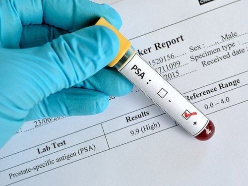
Prostate-specific antigen (PSA) test: The mechanism of this test is to analyze a blood sample for levels of PSA, a protein that the prostate gland produces. prostate created. PSA levels that are higher than normal may indicate an increased risk of cancer.
Digital Rectal Examination (DRE): This test examines the lower rectum and prostate gland to check for abnormalities in size, shape, or texture. The term "digital" means that the doctor uses a gloved, lubricated finger to conduct the examination.
If any abnormalities are detected during the screening, the doctor may perform the following imaging tests:
Ultrasound of the prostate gland : Also known as a transrectal ultrasound, an ultrasound This tone provides an image of the prostate gland and surrounding tissues. An ultrasound is done by inserting an ultrasound probe into the rectum. The transducer sends and receives sound waves through the wall of the rectum into the prostate gland located in front of the rectum. Your doctor may also use ultrasound to guide the needle for the biopsy sample.

MRI of the prostate: An MRI is a way of using a strong magnetic field, radiofrequency pulses, and a computer to create detailed images of organs, soft tissues, bones, and virtually all lateral structures. in the body. The doctor can examine the images on a computer screen, transfer and print, or copy the images to a CD. MRI does not use radiation (x-rays) and MRI is also used to determine if cancer has spread to nearby lymph nodes or bones.
Mp-MRI Prostate: Multisymmetric MRI is a new advanced imaging test. It is a combination of three MRI techniques to provide information about the structure and function of the prostate gland.
Biopsy: This procedure is done by taking a small amount of tissue with a needle from several areas in the prostate gland.
MR or Mp-MRI ultrasound can be used to guide the needle during biopsy.
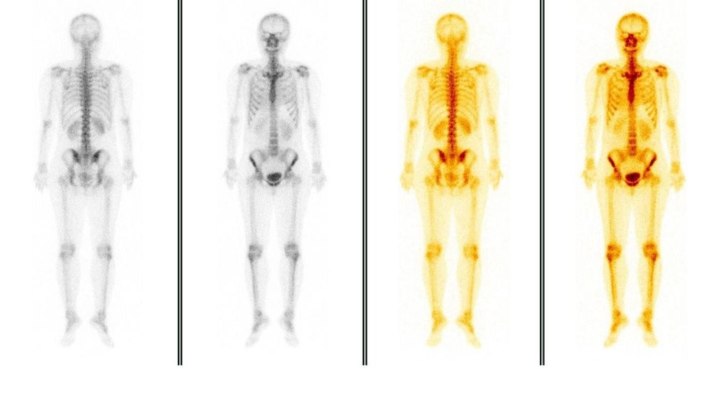
Bone scan : Your doctor may perform a bone scan to determine if cancer has spread to the bones. During a bone scan, a small amount of radioactive material is injected into the blood. When the radiometer passes through the area to be tested. It will emit radiation in the form of gamma rays and the gamma camera will record that image. This information is then transferred to a computer that creates an image of the patient's bones.

PET/CT : A PET/CT scan used to see if prostate cancer has come back (recurred). Like a bone scan, PET/CT inserts a radiometer into a blood vessel. Radioactive sensors attach to proteins on the surface of prostate cancer cells or are taken up by the cancer cells for metabolism.
Vinmec International General Hospital is one of the leading prestigious facilities in the country in the field of screening and early detection of gynecological cancers, including ovarian cancer. Vinmec currently has a package of screening and early detection of gynecological cancer with the following advantages:
A team of highly qualified and experienced doctors. Comprehensive professional cooperation with domestic and international hospitals: Singapore, Japan, USA, etc. Comprehensive treatment and care for patients, multi-specialty coordination towards individualizing each patient. Having a full range of specialized facilities to diagnose the disease and stage it before treatment: Endoscopy, CT scan, PET-CT scan, MRI, histopathological diagnosis, gene-cell testing, .. There are full range of main cancer treatment methods: surgery, radiation therapy, chemotherapy, stem cell transplant... If there is a need for further consultation and examination at the Hospitals of the Health system Vinmec nationwide, please book an appointment on the website to be served.
Please dial HOTLINE for more information or register for an appointment HERE. Download MyVinmec app to make appointments faster and to manage your bookings easily.
Reference sources: radiologyinfo.org, slideshare.net


