This is an automatically translated article.
The article was professionally consulted with Master, Doctor Le Anh Viet - Radiologist - Department of Diagnostic Imaging and Nuclear Medicine - Vinmec Times City International Hospital.Acute appendicitis is one of the most common surgical emergencies. Ultrasonography of appendicitis is a common diagnostic method with high accuracy, convenience and reasonable cost. The ultrasound image of appendicitis will help determine the extent of appendicitis.
1. Overview of appendicitis
Appendicitis is a common appendicitis in adults between the ages of 20 and 30. The disease is less common in the elderly and in young children, however, when acquired, complications of the disease increase the risk of death.Acute appendicitis is a blockage and increased pressure in the lumen of the appendix, creating conditions for bacteria to grow, invade the appendix wall and lead to acute appendicitis. The disease can cause complications of necrosis and perforation of the appendix, so when suffering from abdominal pain due to acute appendicitis, the patient is often taken to the hospital in a surgical emergency.
Most obstructions in the lumen of the appendix cause acute appendicitis, in which obstruction can be caused by:
Fecal stones Enlarged submucosal lymph cysts Foreign body Tumors excess or cecum.
2. Signs of appendicitis
Clinically, acute appendicitis has the following signs:Initially, the pain appears in the epigastrium and around the navel, then spreads to the right iliac fossa. In this position, the patient feels constant, dull pain with increasing intensity. There may or may not be nausea and vomiting. There may or may not be constipation or diarrhea. Loss of appetite May be normal or mild fever. In case the patient has a high fever, there may be complications of appendicitis perforation.

Đau vùng hố chậu phải có thể là dấu hiệu của viêm ruột thừa
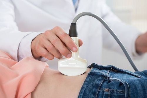
3. Ultrasonography of acute appendicitis
Ultrasound is a common method used in the diagnosis of appendicitis because of its fast, convenient, non-radioactive and low cost.Ultrasound is a method to help detect appendicitis with sensitivity, specificity and positive predictive value of up to 98%.
3.1 Appendicitis on ultrasound Image of appendix is normal
Normally, the rate of appendix ultrasound that can be seen in adults is from 2 to 35% and in children it is 50%. The normal appendix is less than 6mm in diameter, and when compressed, the diameter is reduced to less than 5mm.
On ultrasound, the cross section of the appendix has a target-like appearance with alternating concentric rings with thick and thin echoes. The longitudinal section of the appendix is finger-shaped.
When performing the compression technique, the appendix is only partially collapsed, not completely collapsed like the small bowel loop, and there is no peristalsis at all. The appendix has one end, while the small intestine has two open ends.
Image of acute appendicitis alone
Ultrasonography of acute appendicitis shows the following features in the appendix:
The diameter of the appendix is more than 6mm, the wall of the appendix is more than 3mm thick. The boundaries between the layers are not clear due to inflammation. Pressure does not collapse, sharp pain when pressed. Gallstones can be found in the appendix. Appendiceal stones cause echogenicity, have a dorsal shadow, and are often located at the base of the appendix. Doppler ultrasound showed increased color signal in the submucosa because the blood vessels of the appendix were located mainly in the submucosa. Ultrasonography of acute appendicitis shows the following extra-appendiceal features: Periappendiceal lymph nodes. The cecum wall thickened near the site of the inflamed appendix. The terminal ileum septum is thickened due to diffuse inflammation. The right iliac fossa should have free fluid. Inflammation of the right iliac fossa can cause functional ileus or small bowel obstruction due to edema of the terminal ileum. However, functional ileus or small bowel obstruction is common in complicated appendicitis. Image of necrotizing appendicitis
Ultrasonography of acute appendicitis with necrotic complications shows the following characteristics:
Uneven appendiceal wall thickening, hypoechoic, or thin appendix wall due to purulent-filled interior .. Loss of class structure. Around the appendix is infiltrated fat. Normally, the echo is poor, however, when the echogenic infiltrates are thick, the background contrasts with the poor echo image of necrotizing appendicitis. Doppler ultrasound showed no color signal in the submucosa. Image of appendicitis complicated by abscess
Ultrasonography of acute appendicitis complicated by abscess shows the following characteristics:
Appendicitis abscess can form under the diaphragm, liver, between bowel loops, around the intestines. appendix or pouch of Douglas when the appendix ruptures. The fluid enhances the sound behind the abscess, if it is purulent, there is no echo or echoes within the fluid, in some cases, it is shown above or in the purulent fluid of the abscess with bubbles. gas. Doppler ultrasonography shows congestive signal in the abscess wall. Appendiceal stones can be seen inside the abscess (the right iliac fossa abscess can be of the appendix, or it can also be formed by Crohn's disease, perforated cecal carcinoma or cecal diverticulitis, ...) . 3.2 Difficulties in ultrasonography of acute appendicitis The following cases may give false negative results when ultrasound diagnosis of appendicitis :
Patient has obesity that interferes with ultrasound waves. Patients with small bowel obstruction or paralytic ileus (common in patients with complicated acute appendicitis) limited the study. The patient has an abdominal wall reaction, making it difficult to perform the compression technique. The patient is a woman who is more than 6 months pregnant or has a urinary disease that causes the bladder to distended, making it difficult to perform the compression technique. The patient has an abnormal appendix position. The doctor performing ultrasound for acute appendicitis was inexperienced.
Siêu âm là phương pháp giúp phát hiện tình trạng ruột thừa bị viêm với độ nhạy, độ đặc hiệu và giá trị tiên đoán dương tính lên đến trên 98%
4. Is acute appendicitis dangerous?
Acute appendicitis, if not treated promptly and surgically, can lead to dangerous complications such as:Total peritonitis: When the appendix bursts into the abdomen, it can cause systemic infection. With severe severity, the patient has pain throughout the abdomen and abdominal wall reaction, intestinal paralysis causing abdominal distension, bowel obstruction. Appendicitis Abscess: The patient had appendicitis leading to ruptured appendix, however, the omentum and surrounding loops of bowel formed a focal barrier to prevent the inflammation from spreading to the abdomen. At this time, the patient still has symptoms of pain in the right iliac fossa and has a high fever. Examination and performance reveal a smooth, non-movable mass in the right iliac fossa. Pressing on this area causes pain and tension. Blood tests showed an elevated white blood cell count. When the appendiceal abscess ruptures into the abdomen, it can cause peritonitis complications. 2. Appendiceal masses: The patient has appendicitis, but due to good resistance, the omentum and bowel loops are closed. appendix, limiting the progression of the disease. The patient is still in pain but only has a mild fever. On examination, a firm and immobile mass was found in the right iliac fossa, which was mildly painful when pressed and there was no response to the abdominal wall. Laboratory tests show that the white blood cell count may decrease to near-normal levels. Appendiceal masses may progress to dissolving or form an appendix abscess. The image of appendicitis on ultrasound shows the extent of damage to the appendix as simple inflammation, necrosis or abscess.
Ultrasound is a non-traumatic imaging method that can be performed quickly in all medical facilities. The image of appendicitis on ultrasound helps doctors diagnose and determine the stage of the disease for effective treatment. In addition, ultrasonography is also useful in the differential diagnosis of other gastrointestinal diseases such as ileocecal inflammation, mesenteric lymphadenitis, cecal diverticulitis, and gynecological or urological diseases.
Siêu âm là phương pháp chẩn đoán hình ảnh không sang chấn, có khả năng thực hiện nhanh chóng tại tất cả các cơ sở y tế
Please dial HOTLINE for more information or register for an appointment HERE. Download MyVinmec app to make appointments faster and to manage your bookings easily.