This is an automatically translated article.
The article was professionally consulted with Master, Doctor Trinh Van Dong - Radiologist - Department of Diagnostic Imaging - Vinmec Ha Long International Hospital.Breast ultrasound is a safe, effective, and widely used technique to detect breast abnormalities. When a breast tumor is benign, there are some features that are shown on ultrasound to help the doctor make a differential diagnosis.
1. The role of breast ultrasound in detecting breast diseases
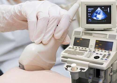
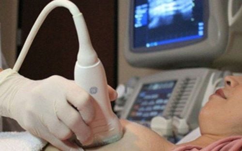
Breast ultrasound is a very commonly used technique to detect breast diseases. Breast ultrasound will help doctors detect abnormalities in breast areas where tumors are suspected and differentiate between fluid-filled cysts or solid masses, benign or potentially malignant breast tumors. Besides, breast ultrasound also helps detect small lesions in the breast that normally cannot be seen or felt.
Another advantage, is that breast ultrasound helps identify mammary gland abnormalities in pregnant women, when other imaging techniques are not available.
Siêu âm vú sẽ giúp các bác sĩ phát hiện những bất thường ở những khu vực vú nghi ngờ có khối u
2. Benign breast ultrasound features
Touching a lump, feeling pain in the breast area is the worry of many women. However, the majority of breast lumps are benign. On the ultrasound image of the breast, the features of benign breast ultrasound include:
The tumor margin is clear and soft, with a thin crust Hyperechoic, homonymous or slightly hypoechoic compared with the normal mammary parenchyma Usually elliptical, in which the major axis is parallel to the skin surface Tumor causes contraction of adjacent structures No signs suggestive of malignancy Some common benign breast tumors in clinical practice such as:
2.1. A mammary gland cyst is a water-filled cyst in the breast that is a benign, painless breast tumor. To the touch, it is felt as a firm, smooth, and movable mass under the skin. Breast cysts are common in women between the ages of 35 and 50, and are rare in women after the age of 50.
Breast cysts have many different sizes, from very small to large sizes 2.5-5cm in diameter. When the breast cyst grows to a large size, it can compress the surrounding tissue, causing an uncomfortable feeling of tightness.
On ultrasound, mammary gland cysts are divided into 3 types with the following characteristics:
Type 1 (simple cyst): On ultrasound images, cysts are round or oval in shape, have clear margins, are empty, and increase. morning after. Type 2 (complicated cyst): Internally, there is a leveled acoustic structure, and other features are similar to simple cysts. Type 3 (complex cyst): On ultrasound, the cyst is thick-walled, thick-walled, and (or) has a solid internal structure.

Nang tuyến vú là một nang chứa nước ở trong vú
2.2. Cystic fibrosis of the breast is a benign breast tumor, manifesting in the ductal system of the lobes and lobules of the mammary gland. The main cause of the disease is that the mammary gland is affected by the imbalance of the hormones estrogen and progesterone. Cystic fibrosis of the breast is common in women 35-45 years old.
The image of cystic fibrosis on ultrasound is very variable, depending on the extent and stage of the lesion:
In the early stages of the disease, although the patient can feel tender tenderness, it is palpable on its own. breast lump, but the ultrasound image may be completely normal. In the late stages: ultrasonography may show cysts, microcalcifications, solid masses or many separate masses. 2.3. Fibrocystic breast fibroadenoma, also known as fibroadenoma, is a benign, non-dangerous growth. Some women have fibroadenoma but have no symptoms while most women will have symptoms such as swelling in the breast, thicker breast tissue, more tender breasts with pain, sensation of discharge. presence of hard mass in one or both breasts. About 10-20% of patients have multiple fibroids and 4% of cases have fibroids in both breasts.
At breast ultrasound, fibroadenomas are characterized by homogeneous acoustic structure, hypoechoic compared with mammary parenchyma, clear margin, coarse calcification or internal wall.
In the case of heterogeneous fibroids, it is necessary to differentiate breast fibroadenoma from medullary, papillary, or mucinous carcinoma.
2.4. Lymphoma of the breast is rare, slow-growing, most of the breast tumors are benign, but a small percentage can be malignant. Lymphoma is a well-defined tumor, composed of connective tissue and epithelial components, the size of which can grow so large that it occupies the entire mammary gland. Tumor usually occurs in the perimenopausal age, over 40 years old.
On ultrasound images, syphilis is usually round or oval in shape. In the case of malignancies, there may be irregular margins, heterogeneous acoustic structure, often hypoechoic, no internal calcification, and posterior wall enhancement.

U mỡ là một khối tế bào mỡ tăng sinh lành tính, không đáng lo ngại
2.5. Lipoma A lipoma is a benign proliferative mass of fat cells that is not of concern. Lipomas are characterized by a soft, painless mass that is well mobile when rolling back and forth with the hand. On ultrasound imaging, lipoma is a well-defined tumor with thin shell, echogenic structure resembling adipose tissue.
All women with risk factors, breast tenderness or palpable breast mass, abnormal nipple discharge... should seek immediate medical attention for timely diagnosis and treatment.
At Vinmec, diagnostic mammography is done carefully and methodically, so a breast ultrasound can last 15-20 minutes.
Customers receive ultrasound and consultation by highly qualified doctors with many years of experience and well-trained in major domestic and foreign medical facilities. Along with making an appointment without having to wait, customers can completely trust and feel secure in choosing Vinmec to examine breast diseases.
Please dial HOTLINE for more information or register for an appointment HERE. Download MyVinmec app to make appointments faster and to manage your bookings easily.