This is an automatically translated article.
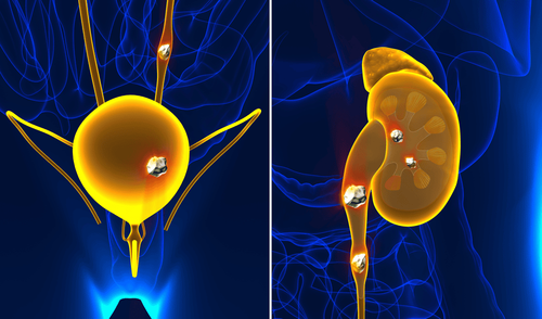
The article was written by MSc Hoang Lan Van Duc - Doctor of Radiology - Department of Diagnostic Imaging and Nuclear Medicine - Vinmec Times City International HospitalDiagnosis of kidney stones should pay attention to the characteristics of size, number, location (kidneys, ureters, bladder, urethra, ..), morphological features (smooth edges, spines, coral shape). ,..), and chemical composition of kidney stones. It helps in the treatment and prognosis of recurrence.
A.The chemical composition of kidney stones
The chemical composition of kidney stones is classified into 5 main types:
Calcium stones: accounting for 60-80% of urinary tract stones Struvite stones (or Magnesium ammonium phosphate): due to bacterial infections, accounting for 5-15% of uric acid stones: accounts for 1-20% Cystine stones: 1-2% Some rare stones (xanthine stones, silica stones, ..): are the result of some rare metabolic diseases, either due to drug use, or due to diet Eat >>> What are the types of kidney stones and which are the most common?

Sỏi thường tồn tại dưới dạng hỗn hợp đan xen giữa các thành phần hoá học
1. Calcium stones
The most common (accounting for 60-80%), including two types of calcium oxalate and calcium phosphate.
Calcium oxalate stones have calcium oxalate as the main ingredient, which can be combined with calcium phosphate. Calcium oxalate stones are dark brown, thorny, very solid, with clear contrast, more common in men than in women.
Calcium phosphate stones in the form of brusite or apatite, ivory white, with many concentric layers, friable, often large in size, clearly contrasted, and equally common in men and women.
Approximately 80-85% of patients with calcium stones do not have an underlying predisposing condition for calculi and are referred to as primary calcium stone disease. About 15-20% of patients with calcium stones have underlying disease prone to stone formation, that is primary hyperparathyroidism, hyperoxaluria, congenital oxaluria, renal tubular acidosis, cushing's syndrome, treatment. prolonged steroids, vitamin D toxicity, prolonged immobilization, milk-alkali syndrome, spongiform nephropathy. This type of calcium stone disease is called secondary calcium stone disease. In addition, decreased urine volume and decreased excretion of citrate and magnesium also increase the risk of stone formation.
2. struvite gravel
Struvite stones, or stones caused by infection, chemical composition is Magnesium ammonium phosphate, accounting for 5-15%.
Pebbles are usually large in size, coral-shaped, ivory-white, solid, and opaque. The rate of the disease in women is twice that of men, corresponding to the rate of urinary tract infections in women is twice that of men. Urinary tract infections in children, occur equally in both sexes, and are often associated with urinary tract abnormalities.
Chronic urinary tract infections in young people, often due to nerve bladder damage, paralysis of the lower extremities, prolonged immobilization after accidents, sexual activity and pregnancy are also risk factors for urinary tract infections. urinary tract in women. Not all urinary tract infections carry a risk of stone formation. Only certain bacteria capable of breaking down urea to produce ammonium, bicarbonate, and hydroxyl ions, along with alkalinizing urine, are at risk of stone formation. Those bacteria are: Escherichia Coli, Proteus, Krebsiella, Streptococcus, Staphylococcus, Pseudomonas.
Usually combined with apatite carbonate or calcium carbonate when urine pH is above 7.8, or add ammonium hydrogen urate when both urea-degrading and uric acid bacteria are present.
3. Uric acid stones
The chemical composition includes uric acid and ammonium urate, accounting for 1-20%.The stone is dark brown, solid, usually round, smooth like a pebble, not contrasted on X-ray film, on CT it usually has a density of 100-200 HU (compared to stones with normal calcium > 400 HU) . Uric acid stones, often combined with calcium oxalate, are common in elderly men. European countries and countries with developed industry, high standard of living, have a higher rate of uric acid stones than Asian countries and countries with slow economic development and low living standards.
Approximately 20% of patients with primary gout, and 40% of patients with secondary gout have uric acid stones in the urinary tract. Risk factors for uric acid stone formation are increased uric aciduria, low urine pH, and low urine volume.
4. Cystine stones
Cystine stones are often caused by hereditary increased amino acid cysteine in the urine, accounting for 1-2%.
The gravel is light yellow or ivory white, solid, and radiopaque. Cystine stones are often associated with calcium phosphate (apatite) stones, which are common in young people.
B. The role of dual-energy CT (DECT) in the assessment of kidney stone composition
Introduction to dual-energy CT (DECT) or spectral CT (GSI):
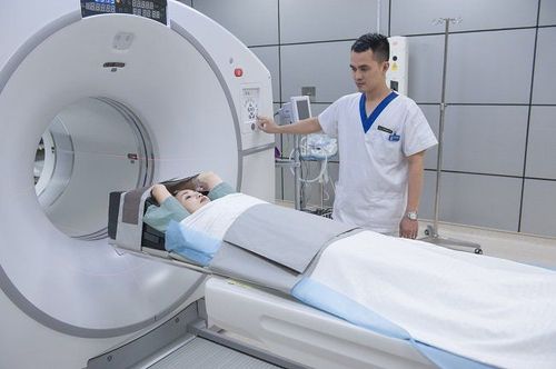
Conventional computed tomography (or single-energy CT) is an invaluable tool in the diagnosis of kidney stones. However, conventional CT has some limitations in analyzing the composition of stones, mainly based on the value of density (unit: HU). Therefore, finding a method that can determine the composition of stones can help patients better treat this pathology. This is especially important when choosing between medical treatment, surgery or interventional procedures, such as possibly medical treatment of uric acid stones. Recently, dual-energy CT (DECT) or spectral CT (GSI) has been shown to be useful in the evaluation of kidney stones.
At Vinmec Times City International Hospital, equipped with GE's latest CT scanner, with the principle of two energy levels generated from one source, there is a quick transition between high (140 kVp) and low energy levels. (80kVp) is only about 0.5 milliseconds. That acquired data is processed and converted into different energy spectra. The attenuation of X-ray energy as it passes through a body tissue measured at different energies (high and low), which can be transformed into an attenuation measurement as the density (or quantity) of two objects. whether necessary to produce a measurable deterioration. This process, known as material pair separation, may offer the possibility of tissue characterization. Materials with low (water) and high (iodine) attenuation were chosen as the base pair, in addition to many other dose pairs, even separating the 3 matter.
From the obtained data, the effective atomic number (Z) of that tissue's constituent material can be inferred, or the composition of body tissue can be distinguished. Furthermore, through the reproduction of monochromatic images, it is possible to minimize image noise in the examination area due to beam hardening.
Please dial HOTLINE for more information or register for an appointment HERE. Download MyVinmec app to make appointments faster and to manage your bookings easily.
C. Principle of determining the gravel composition of two-energy CT
1. Based on comparison of gravel densities at low and high energies
With this method, we measure the average density of the stone at a low energy level, and then compare it with a high energy level.
According to some studies such as Hidas in 2010, Dawoud in 2017 evaluated the stone composition on DECT compared with the crystallographic analysis of stones, it was possible to distinguish 3 types of stones: calcium stones, uric acid stones and stones. cystine, specifically: uric acid stones have a density of about 325-550 HU at low energy levels, about 300-550 HU at high energy levels. Cystine stones have a density of about 1000-1800 HU at low energy levels, about 900-1500 HU at high energy levels. Calcium stones have a density of 650-1900 HU at low energy levels, about 450 - 1350 HU at high energy levels.
The low/high energy HU ratio if < 1.1, suggests uric acid stones, 1.1-1.24 suggests cystine stones, 1.25-2.4 suggests calcium stones.
However, the disadvantage of this method is that it cannot distinguish calcium oxalate stones from calcium phosphate stones, and cannot distinguish between struvite stones and calcium stones because they have the same energy loss rate as calcium depletion. , the composition of mixed stones could not be determined (especially stones combining uric acid with hydroxy apatite).
2. Based on the separation of matter pairs and determine the effective atomic number (Z number) of matter
The data obtained when taking two-energy CT scans will be processed with material pair separation, in determining kidney stone composition often use calcium and water, calcium and uric acid material pairs, creating monochromatic images. with calcium or uric acid removal information. With this method, it is possible to distinguish uric acid stones from non-uric acid stones.Uric acid stones will be clearly visible on the calcium removal image, no color overlay on the calcium color-coded image, and no longer clearly visible on the uric acid removal image. As for non-uric acid stones, on the contrary, it is not clear on the calcium removal image, there is color coating on the calcium color image, clearly observed on the uric acid removal image.

Hình 2: ca 1. (a) ảnh gốc cho thấy sỏi ở vị trí đài giữa thận phải, (b) tái tạo bản đồ màu canxi, có màu canxi ở vị trí sỏi, (c) ảnh xóa axit uric thấy rõ hình sỏi, (d) ảnh xóa canxi thấy mờ hình sỏi, chứng tỏ thành phần sỏi có canxi

Hình 3: ca 2. (a) ảnh gốc cho thấy sỏi ở vị trí chỗ nối bể thận – niệu quản trái, (b) tái tạo bản đồ màu canxi, không thấy phủ màu canxi ở sỏi, (c) ảnh xóa canxi thấy còn tồn tại sỏi, (d) ảnh xóa axit uric thấy mất hình sỏi, chứng tỏ thành phần sỏi là axit uric

Hình 4: (ca 1). Hình phải: ảnh gốc với ROI đo số nguyên tử hiệu dụng Z. Hình trái: ảnh phổ cho thấy Z trung bình của sỏi có giá trị từ 12 – 15 chỉ ra rằng thành phần của sỏi có canxi.

Hình 5: (ca 2): Hình phải: ảnh gốc với ROI đo số nguyên tử hiệu dụng Z. Hình trái: ảnh phổ cho thấy Z trung bình của sỏi có giá trị từ 6.9 – 9 chỉ ra rằng thành phần của sỏi có axit uric
Please dial HOTLINE for more information or register for an appointment HERE. Download MyVinmec app to make appointments faster and to manage your bookings easily.
References to the article:
Chaytor R.J., Rajbabu K., Jones P.A., et al. (2016). Determining the composition of urinary tract calculi using stone-targeted dual-energy CT: evaluation of a low-dose scanning protocol in a clinical environment. Br J Radiol , 89 ( 1067 ), 20160408. Dawoud M.M., Dewan K.A.A.W.A., Zaki S.A., et al. (2017). Role of dual energy computed tomography in management of different renal stones. Egypt J Radiol Nucl Med , 48 ( 3 ), 717–727. Hidas G., Eliahou R., Duvdevani M., et al. (2010). Determination of Renal Stone Composition with Dual-Energy CT: In Vivo Analysis and Comparison with X-ray Diffraction. Radiology , 257 ( 2 ), 394–401. Kriegshauser J.S., Paden R.G., He M., et al. (2018). Rapid kV-switching single-source dual-energy CT ex vivo renal calculi characterization using a multiparametric approach: refining parameters on an expanded dataset. Abdom Radiol , 43 ( 6 ), 1439–1445. Kulkarni N.M., Eisner B.H., Pinho D.F., et al. (two thousand and thirteen). Determination of Renal Stone Composition in Phantom and Patients Using Single-Source Dual-Energy Computed Tomography:. J Comput Assist Tomogr , 37 ( 1 ), 37–45. Leng S., Huang A., Cardona J.M., et al. (2016). Dual-Energy CT for Quantification of Urinary Stone Composition in Mixed Stones: A Phantom Study. Am J Roentgenol , 207 (2) , 321–329. Vasilyeva E. (2011). Dual Energy CT (DECT) for determination of renal stones composition before extracorporeal shock-wave lithotripsy (ESWL). 1–24.