
This is an automatically translated article.
Article written by MSc Vu Duy Dung - Department of General Internal Medicine - Vinmec Times City International Hospital
Tuberculomas may occur in the brain, spinal cord or subarachnoid space, subdural or epidural and are often accompanied by periphery edema and ring enhancement.
1. Tuberculosis (tuberculous granuloma)
Ten percent of patients with tuberculous meningitis also have tuberculous tumors; with a third of such patients having multiple tuberculosis. The clinical presentation of CNS TB varies with the location of the lesion but usually includes headache, seizures, focal neurological deficits, and papilledema.
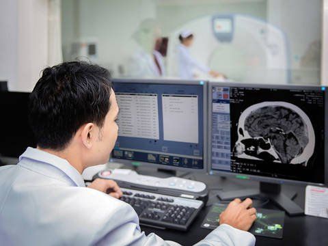
On neuroimaging, tuberculous tumors are usually space-occupying lesions; Because the center of the tubercle is necrotic and liquefied, the neuroimaging features on T2 and FLAIR pulsed MRI vary from hypointensity to co-signaling to hyperintensity. The neuroimaging features of CNS TB vary with the age of the lesion and are not specific for CNS TB.
While in adults, TB tumors are usually above the tent, in children the lesions are usually below the tent. The CSF of patients with TB is often indistinct, while TB skin tests are positive in 85% and chest radiographs are abnormal in 30% to 80% of cases.

U lao có thể xảy ra ở cả người lớn và trẻ em
2. Tuberculosis abscess
Tuberculosis abscesses are seen in less than 10% of patients with CNS TB and likely represent a later stage of tuberculosis that occurs after granulation; An abscess contains more bacilli than a granuloma.
Clinical symptoms caused by a tuberculous abscess usually include fever, headache, and focal neurological deficits. Differentiation between tuberculous tuberculoma and tuberculous abscess can be difficult on neuroimaging, but an abscess wall tends to be thicker, may be divided into several compartments, and is often strongly enhanced. The following clinical case illustrates an insidious presentation of a tuberculous CNS abscess.
On diffusion pulses (DWI), MRI may show limited diffusion in tuberculosis or abscesses with fluidized necrosis but, in general, this finding is absent in only solid necrotic lesions. .
Clinical case:
43-year-old male came to the clinic for a second opinion on neurology. Seven months ago the patient presented to an emergency department with symptomatic aphasia and progressive somnolence, and brain MRI revealed a mass in the left frontal lobe with heterogeneous enhancement and mild surrounding edema.
Diagnosis is follow-up glioma , and the patient was biopsied, the results showed no diagnosis. The patient's speech continued to worsen, and 1 month later he developed right hemiplegia. Re-evaluation revealed a heterogeneously enhanced left frontal lobe mass of 5cm x 7cm. The patient underwent complete surgical resection of the brain lesion.
Neuropathology showed no evidence of glioma or lymphoma; Gram stain, methenamine-silver stain, and acid-fast stain showed no microorganisms.

Áp xe lao gặp trong ít hơn 10% bệnh nhân mắc bệnh lao hệ thần kinh trung ương
On examination, patients often find it difficult to find words and cannot name a straw or paper clip. The patient has a drooping right lower half of the face, and hearing loss in the left ear. The patient has a mild right hemiplegia and asymmetrical tendon reflexes (the right side is more sensitive than the left). Sensation with pins decreased all over the right half of the body.
Chest CT showed calcified granulomas in the lung and mediastinal lymph nodes, consistent with prior granulomatous disease. Examination of a paraffin block of brain parenchyma taken from the primary biopsy sent back to the molecular microbiology laboratory for PCR testing of mycobacteria and mycobacterial DNA sequencing results consistent with CNS tuberculosis nurse. Follow-up MRI after 3 months of 4-drug anti-TB treatment showed heterogeneous enhancement that had turned into ring-shaped enhancement. Edema around the lesion remains.
After 18 months of antituberculosis therapy, neurologic examination improved muscle strength to normal in all extremities without proximal deformity of the joints, and speech was rather hesitant but fluent. Brain MRI continued to show improvement, with mild enhancement. After 12 months following anti-tuberculosis therapy, MRI showed no enhancement and edema, although residual encephalomalacia remained.
This case demonstrates the difficulty of diagnosing tuberculous abscess on neuroimaging, the often time lag between pre-existing pulmonary TB and CNS TB development, and the usefulness of laboratory testing. Advanced diagnostics, in this case PCR mycobacteria, for the diagnosis of tuberculous granulomas. The majority of TB cases in the US are in people born outside the US.
Chẩn đoán áp xe lao trên hình ảnh thần kinh thường gặp nhiều khó khăn
3. Tuberculous myelitis
Tuberculous myelitis is an uncommon complication of TB infection that can occur as the first manifestation of CNS TB, as a spread of TB infection from one vertebra to the next contiguous, or as a complication of tuberculous meningitis.
Tuberculous radiculopathy often causes lower extremity weakness, is progressive, subacute with bladder dysfunction, paresthesia, radicular pain, and muscle atrophy. The neuroimaging of tuberculous radiculitis often shows opacification of the spinal subarachnoid space, loss of spinal cord boundaries, fusion of nerve roots, and increased nodular intradural enhancement.
Although there is no randomized controlled trial of steroid therapy for tuberculous myelitis, most experts recommend concurrent administration of steroids with antituberculous therapy.
Vinmec International General Hospital is one of the hospitals that not only ensures professional quality with a team of leading medical doctors, a system of modern equipment and technology. The hospital provides comprehensive and professional medical examination, consultation and treatment services, with a civilized, polite, safe and sterile medical examination and treatment space.
Please dial HOTLINE for more information or register for an appointment HERE. Download MyVinmec app to make appointments faster and to manage your bookings easily.