This is an automatically translated article.
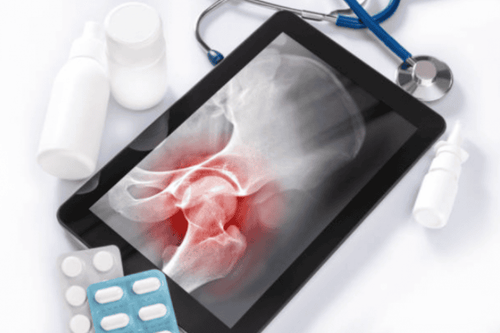
The article was professionally consulted by Specialist Doctor I Vo Cong Hien - Radiologist - Radiology Department - Vinmec Nha Trang International General Hospital.Injecting pain relief directly into the joint is a quick method of pain relief but can bring many disadvantages, especially with small joints, difficult to achieve high accuracy. Therefore, it is necessary to have the support of guiding means such as using enhanced radiography.
1. Injection of joint pain relief under enhanced X-ray
X-ray is one of the oldest techniques but is still frequently indicated in diagnostic imaging, especially in musculoskeletal diseases. X-ray is an imaging technique that uses X-ray radiation to pass through the examination body in a small enough dose and create images of the internal organs of the body. Joint X-rays help doctors assess changes in joint structure and function and help determine possible treatment requirements, including surgery or knee replacement. With TV-enhanced X-ray, the image created is clearer and brighter than conventional X-ray machines, helping doctors see more clearly and in detail to make key conclusions. body for the patient.Usually, when performing joint pain relief injections, the doctor only needs to insert the needle directly without the need for a guide vehicle. However, in difficult cases, deep small joints need guiding means such as enhanced X-ray to ensure accuracy and safety for the patient. In addition, under the guidance of enhanced X-ray, it allows the doctor to monitor the drug's spread in the joint as well as the drug outflow (if any).
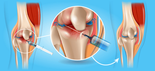
Tiêm giảm đau khớp sử dụng X quang tăng sáng giúp bác sĩ theo dõi sự lan tỏa của thuốc trong ổ khớp
2. Arthritis pain relief injection technique under enhanced X-ray
Before starting to perform the technique, the patient is thoroughly explained about the procedure to coordinate with the doctor. In addition, the patient may also be asked to fast before 6 hours. Can drink no more than 50ml of water. In the intervention room: the patient lies on his stomach, back or side depending on the device to monitor breathing, pulse, blood pressure, electrocardiogram, SpO2. Disinfect the skin, then cover with a sterile, perforated cloth. In case the patient is too excited, can't lie still, the doctor can give him a sedative.Starting the procedure, the patient is placed on the projector table, disinfecting the skin of the joint to be poked. The doctor washes his hands, wears a shirt, wears gloves, and a sterile sheet. Next, the doctor will locate the joint space to poke. Insert the needle into the joint socket. Inject 1ml of contrast agent to confirm the position of the needle. Inject the drug suspension in the ratio of 1.5ml Depo-Medrol 40mg + 1ml Lidocaine 2% + 1ml contrast agent, the volume depends on the joint position to be injected. After injecting the drug, the doctor will monitor the spread of the drug under the light curtain.
The procedure is considered successful when it ensures that the needle is in the joint, the drug mixture is diffused in the joint, and the pain relief effect is right after the procedure.
3. Complications can be encountered when injecting joint pain relief under bright X-ray
Bleeding at the puncture site: If the patient notices bleeding, notify the medical staff so that the puncture site can be bandaged. Soft tissue hematoma next to the needle puncture site: in this case, the patient will continue to be monitored. Joint infection Injection therapy for joint pain under enhanced X-ray is a technique that requires a high level of experience of the doctor and the perfect coordination of the patient. To achieve high diagnostic efficiency, patients need to choose reputable addresses with x-ray machines and modern and standard medical equipment.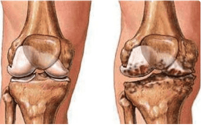
Nhiễm trùng khớp có thể xảy ra khi tiêm giảm đau khớp dưới X quang tăng sáng
BSCK I Vo Cong Hien has many years of experience in the field of imaging, especially in diagnostic ultrasound for general abdominal diseases, advanced cardiac - vascular, obstetrics and gynecology. Doctor Vo Cong Hien used to work at the Imaging Department of the University of Medicine and Pharmacy Hospital - Hoang Anh Gia Lai before working at the Imaging Department of Vinmec Nha Trang International General Hospital.
Customers can directly go to Vinmec Health system nationwide to visit or contact the hotline here for support.