This is an automatically translated article.
The article is professionally consulted by Master, Doctor Nguyen Quang Duc - Doctor of Nuclear Medicine - Department of Diagnostic Imaging and Nuclear Medicine - Vinmec Times City International Hospital. The doctor has more than 12 years of experience in the field of Diagnostic Imaging.There are different types of radioisotopes, each of which tends to be concentrated in different organ tissues. Therefore, the type of radioisotope used depends on the part of the body to be imaged. Gallium scans tend to focus on areas of the body with rapid cell division. Areas with high concentrations of gallium can be suggestive of infection, inflammation, injury, or cancer.
1. What is a Gallium scan?
A Gallium scan is a nuclear medicine procedure that can examine problem areas in certain tissues of the body. In a gallium scan, a small dose of a radioisotope is given intravenously. This radioactive isotope will emit a type of light that is invisible to the naked eye, called gamma rays. Accordingly, a special type of camera, a gamma camera, can detect this type of ray. Use this camera to capture gamma rays after radioactive isotope injection.2. Gallium scintigraphy mechanism of action
The radioactive tracer gallium citrate will be injected into a vein in your arm. This substance has iron-like activity, some will bind to proteins in the blood, especially a protein called transferrin. It is then transported through the bloodstream and begins to accumulate in different areas of the body. These tissues include bone tissue, liver, intestines, and areas of inflammation or places where white blood cells accumulate.Gallium accumulation can take several days, which explains why the second part of the scan is done a few days later. After the tracer builds up in the body, the gamma camera takes pictures of the gamma rays emitted from inside the body, which are then converted into electrical signals and transmitted to a computer. The computer will generate images with different colors or grayscales corresponding to the received signals of different intensities. The photos show areas with a higher than normal amount of tracer known as hot spots.
For example, areas of target tissue that emit a lot of gamma rays are represented as red areas on computer images. Areas of target tissue that emit low gamma rays are shown in blue (cold areas). Various other colors can be used in the “middle range” depending on the level of gamma rays emitted.
3. The role of the Gallium scintigraphy

4. Gallium scintigraphy process
Gallium scans are usually done by a nuclear medicine technician. In most cases, a radiologist or nuclear medicine specialist will interpret the images.Scanning technique is done in 2 parts. The first part is intravenous gallium citrate. After the injection, the patient is usually sent home so that the gallium can circulate in the body. The second part of the technique is taking pictures. Depending on the hospital and the reason for the scan, the patient may be instructed to return to the hospital for imaging 1 day, 2 days or 3 days after the injection.
When you arrive for the scan, you will need to remove your jewelry, which may require removing all or most of your clothing depending on the area being inspected.
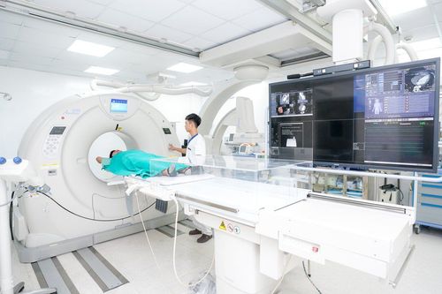
With the patient lying on his back on a table, a large nearby camera scans the radiation released by the tracer and creates an image of the tracer in the tissues. The camera may move slowly over and around the body. The camera does not produce any radiation, so the patient does not need to worry about being exposed to more radiation while the scan is being done.
Note that the patient needs to lie still during each scan to avoid blurring the image. You may be asked to keep your breath short for some scans. The number of images recorded and the time interval between each image will vary depending on which part is being recorded. Each scan can take about 60 to 90 minutes.

5. Meaning and value of Gallium scan results
A Gallium scan is a nuclear medicine procedure that takes pictures of certain tissues in the body after a radioactive tracer is inserted. Test results are usually ready within 2 days of scanning.Normal: The collection and activity of gallium in the bones, liver, spleen, and colon are normal. No areas of abnormal gallium were seen. Abnormal: An abnormally high amount of gallium (hot spot) is present in one or more areas of the body. This could mean inflammation, infection, or a tumor. Gallium scans are used for certain types of cancer, mainly lymphoma, bone, or bone marrow. However, a negative value does not exclude the possibility of cancer, as some cancers do not appear on a Gallium scan. The technique also cannot tell if a tumor is cancerous (malignant) or noncancerous (benign).
The doctor will explain the results along with the results of other tests (physical examination, blood tests, X-rays...). In many cases, results from an MRI or positron emission tomography (PET) scan can be as accurate as those from a Gallium scan. If other nuclear scanning procedures need to be performed, they should be scheduled before the Gallium scan. This is because the gallium tracer stays in the body longer than other tracer compounds.
6. What to keep in mind when scanning Gallium

Usually, the gallium scan does not cause any side effects. Through natural decay, small amounts of radioactive material in your body lose their radioactivity over time, and are then eliminated from the body in urine or feces during the first few hours or days after surgery. radiography. Patients may be instructed to take special precautions after urinating, flushing the toilet twice and washing hands thoroughly.
If you come into contact with children or pregnant women, tell your doctor. Although the level of radiation used in scintigraphy is very small, special precautions are still needed. The hospital will provide patients with more information on this matter.
If you have to go abroad within 3 months after the scan, ask your doctor for more detailed instructions. Airport doors have very sensitive radiation detectors that can detect very small amounts of radioactive material remaining in the body after a scan. Usually, the hospital will probably provide a confirmation letter that you can show to staff at the airport.
Please dial HOTLINE for more information or register for an appointment HERE. Download MyVinmec app to make appointments faster and to manage your bookings easily.


