This is an automatically translated article.
The article is professionally consulted by Master, Doctor Nguyen Quang Duc - Doctor of Nuclear Medicine - Department of Diagnostic Imaging and Nuclear Medicine - Vinmec Times City International General HospitalIn the field of nuclear medicine, PET CT scanning technique is considered as one of the modern imaging methods and has value in diagnosing as well as monitoring the treatment process of certain diseases. Therefore, it is necessary to understand the basic principles of PET imaging to be able to apply this method correctly and effectively.
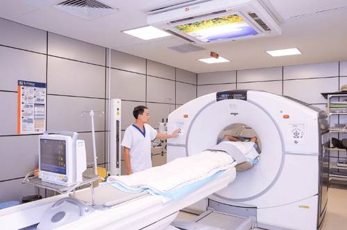
1. PET CT scan
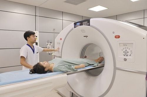
PET CT scan is one of the paraclinical methods that play a huge role in finding tumors in the organs of the body that other imaging methods are difficult to investigate. In addition to the diagnosis, PET CT scans also help doctors monitor the effectiveness of the treatment process on cancer patients, from which there are further treatment directions for the patient to recover quickly. in the shortest time.
PET CT scan allows to survey the anatomical structures of the organs, and also investigate the problems related to the metabolic function of the tumors in those organs, if any. In addition, PET CT can also aid in the study of certain diseases related to the nervous or cardiovascular system, even some infections. A number of studies in the world have shown that thanks to the support of PET CT scanning technique, some treatment indications on cancer patients have changed since then, helping patients have a good treatment result. more than the original.

2. PET imaging principle
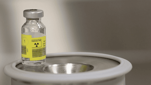
The basic principle of PET imaging is the process of concentrating radiopharmaceuticals on physiological and metabolic grounds in tumors and other benign organ tissues. With the basic physiological processes in the body, the metabolic reactions, metabolic processes as well as protein synthesis in tumors will be higher than in non-tumor organizations and organs. Therefore, the process of transporting and synthesizing amino acids will also take place more in organizations with cancerous tumors. 11C - methionine, 11C - tyrosine are used in PET imaging to detect these hyperactive processes.
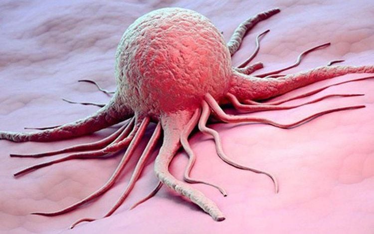
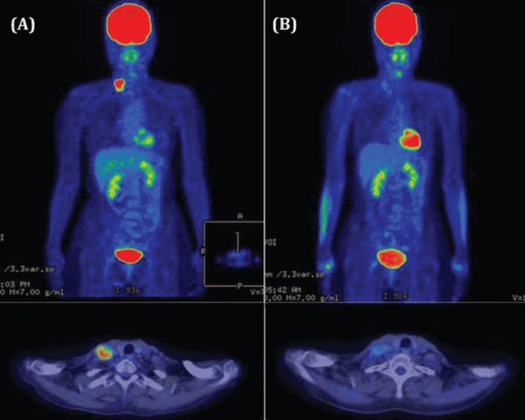
In addition, tumor-forming cells also use glucose to a higher extent than cells in other organs, so 18F with Glucose is used for PET imaging in these cases. . For radiopharmaceuticals, FDG, when moving to cancer cells through the blood and GLU1 transporter, it will undergo phosphorylation under the action of hexokinase and finally form FDG - 6 phosphate. Therefore, by marking the precursors of DNA and Glucose with radioactive substances, when there are tumors appearing in the body, this information will be recorded and reflected in detail about the tumor in the body. anatomically as well as metabolically of these cancer cells.
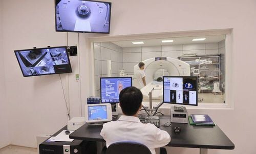
The principle of PET imaging can also be expressed by a number of operations involving positrons. In particular, when there is positron decay, the pair of photons will be released and travel in opposite directions, which will be detected by the detectors placed in it, so that images can be recorded. when this process takes place. Each pair of photons recorded by the detector, also known as a coincident pair, is recorded with a separate encoding, then transmitted to the computer receiving information and using a number of specialized algorithms to process the information. information and give PET CT scan results. PET CT imaging systems have also improved over time, with the introduction of several types of crystalline materials such as BSO, LSO, LYSO..., as well as innovations in probes and new TOF technologies. Regarding the issue of implementation time, modern imaging technologies such as 2D, 3D... can bring the highest efficiency in the detection and treatment of related pathologies. In particular, image reconstruction technology MLEM, OSEM... has reduced the shortcomings of PET CT scanning techniques in the past, such as problems of noise, scattering, resolution... greatly improve image quality. In recent years, the PSF technique has greatly increased the resolution of images when taking PET CT, the contrast has also improved significantly, along with the introduction of 4D recording, making the PET CT imaging when the organ is in motion as well as the recognition of the target is made easier but still very effective.
3. Conclusion
PET CT scan is one of the imaging methods indicated in a number of pathologies such as cancer, cardiovascular... to detect tumors as well as evaluate treatment effectiveness. The principles of PET CT imaging are basic and need to be updated every day to be able to take full advantage of the great benefits that nuclear medicine in general this subclinical approach brings in particular.
Currently, Vinmec International General Hospital has become a leading prestigious address in disease screening with modern techniques, proud to be the first private hospital in Vietnam to deploy this technique. Modern and accurate diagnosis with the most modern 128-sequence PET/CT system in Southeast Asia, for accurate images and short imaging time, the team of doctors are leading experts with high expertise and rich experience. Experience creates trust for customers when experiencing medical examination and treatment services at Vinmec.
If you have a need for consultation and examination at Vinmec Hospitals under the nationwide health system, please book an appointment on the website for service.
Please dial HOTLINE for more information or register for an appointment HERE. Download MyVinmec app to make appointments faster and to manage your bookings easily.


