This is an automatically translated article.
The application of the principles, methods and techniques of physics in medical practice and research has created a scientific and technical revolution in the entire medical field. It is applied in clinical examination and treatment of diseases.
1. What is the role of physics in medicine?
Medical physics is applied in medicine to help prevent, diagnose and treat human diseases. Medical physics can be classified into several subgroups including: medical imaging physics, radiation oncology physics, non-ionizing medical radiation physics, nuclear medicine physics.
Medical physics is studied by medical professionals with specialized training in physics, specializing in the study of the application of physics in health care. The concept of medical physics was first introduced by Félix Vicq d'Azir, a French surgeon in 1778.
The main role of physics in medicine is based on the principles of ion, ultrasonic waves. , laser, X-ray ... in the diagnosis and treatment of many diseases.
2. Applications of physics in medicine
The applications of physics in medicine have been widely applied that many technologies we already know. Applications of physics in medicine include:
X-Rays and CT Scans In radiography the signals are generated from a narrow beam of X-rays that pass across the affected area of interest to produce images on movie. Similarly, cross-sectional X-ray images obtained by repeated scanning are superimposed by digitally erasing the background to produce high-resolution, three-dimensional or quadruple computed tomography (CT) images. dimension to analyze dynamic processes.
CT perfusion : Besides providing reconstructed anatomical images, CT scans can produce higher quality tissue perfusion information through perfusion scans motion. By infusion a contrast agent is used and then repeated imaging of the affected area is performed at 3 - 5 s intervals for 30 s. These images are then superimposed to form 4-D images, which are useful in analyzing hemodynamic parameters, including blood flow and blood volume.
Magnetic Resonance Imaging Magnetic resonance imaging (MRI) is a non-invasive medical imaging technique. Application using static magnetic fields, magnetic gradients and computer-generated radio waves to create high-quality 3-D images of tissues and organs. The magnetic field causes the body to rearrange its protons with that magnetic field. Then, radio waves excite the protons and an MRI sensor is used to detect the energy (signal) released from the protons. In quantitative MRI, the difference in contrast between two tissues is maximized on a single image using the difference in relaxation time of the 2 measured tissues. These images are based on the characteristics of a particular tissue. Commonly used cases for MRI are to measure cerebral blood flow and analyze soft tissue structures.
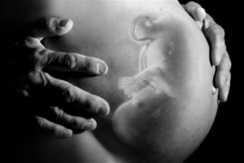
Ultrasound Ultrasound is a high-frequency sound wave that is passed through the body to create non-invasive images of various tissues and organs. Differences in the mechanical properties of different organs or tissues in the body cause ultrasound reflections. These reflections are measured to create an ultrasound image.
The main advantage of ultrasound compared to other medical imaging techniques such as CT and MRI is also good efficiency in many cases, low cost, safety, and ease of implementation. Besides disease diagnosis, ultrasound is used for therapeutic purposes. For example, high-intensity focused ultrasound waves remove affected tissue inside the body without damaging surrounding healthy tissue. In addition, ultrasound is used for targeted injection or aspiration.
However, it will have more errors in diagnosing some diseases. Still need CT and MRI intervention.
Nuclear Medicine In nuclear medicine, radioactive substances are used to observe physiological processes of the body. Radioactive substances are also used to deliver a targeted therapeutic dose. A very small amount of radioactive material is introduced into the body during the procedure. The radioactive material is then absorbed by the organ or tissue under investigation. The radiation emitted by the detector as a result of decomposition is detected by a gamma camera, which generates digital signals to analyze the functional state of the organ.
The gamma camera produces a 2-D image when standing still. In single-proton emission computed tomography, the camera is rotated to produce axial slices of the target organ. These slices can be used in PET-CT scans to create 3-D images. Nuclear medicine is often used to detect a tissue that has absorbed abnormal amounts of radiation, and to detect cancer early.
Radiation Therapy Radiation therapy involves delivering a dose of ionizing radiation inside the body to help destroy and eliminate cancer cells or certain other tumors in the body. During the entire treatment procedure, medical imaging is performed to ensure safe, targeted delivery of radiation and to evaluate radiation-induced changes in anatomy.
In general, thanks to physics, the medical profession has a turning point in advanced science. Scientists continue to research to have more diagnostic and therapeutic methods to be applied in medicine.
Please dial HOTLINE for more information or register for an appointment HERE. Download MyVinmec app to make appointments faster and to manage your bookings easily.
Reference source: news-medical.net


