This is an automatically translated article.
The article is professionally consulted by Master, Doctor Le Hong Chien - Doctor of Radiology - Intervention - Department of Diagnostic Imaging and Nuclear Medicine - Vinmec Times City International General Hospital.An aortic aneurysm is a disease that occurs when the inner lining of the artery is torn, causing blood flow to rapidly peel away the middle layer of the artery wall. This is a medical emergency and imaging plays an important role in the diagnosis and treatment of aortic dissection.
1. Features of aortic dissection
Aortic aneurysm is a rare disease (the incidence of this disease is only 5-30 cases/million people/year), and the frequency depends very much on the variation of each population with other factors. different risk factors.Symptoms of aortic dissection are very diverse and easily confused with many other emergency conditions, so we need to pay attention to different suspicions to be able to accurately diagnose and treat promptly. time. Incidence in men/women ~ 2/1. The age of the disease is highest in the range of 60-70 years old with a mortality rate of 1%/hour in the first 48 hours.
The most common site of injury is the ascending aorta (accounting for ~ 65%), descending aorta (accounting for ~ 20%), arch of the aorta (accounting for ~ 10%), the rest is aortic dissection aneurysm. belly.
Predisposing factors of aortic dissection: high blood pressure (70-90%), collagen and elastic fiber degeneration, trauma, physician-induced complications, aortic stenosis, coarctation motherboard...
2. Classification
Aortic aneurysms are divided into many different types, including:● DeBakey-based classification will be divided into 3 main types such as:
○ Type 1: are lesions of the entire ascending aorta along with arteries motherboard down.
○ Type 2: for lesions appearing only in the ascending aorta.
○ Type 3: lesions appear only in the descending aorta.
● The Stanford classification will be divided into:
○ Type A: lesions in the ascending aorta, regardless of onset in any part of the aorta in the body.
○ Type B: are lesions of the aorta distal from the main origin of the inferior left clavicle groups.
● Classification based on the time that the patient has the disease.
○ Acute: calculated when the patient has been less than 2 weeks since the onset of illness.
○ Chronic: is when the patient's illness is more than 2 weeks, according to research, up to 1/3 of patients when diagnosed and treated are in the chronic group.
○ The mortality rate will rapidly increase within the first 2 weeks, and can peak to about 75-80%, causing a natural threshold for the general course of the disease.
3. The role of imaging in diagnosis and treatment of aortic dissection
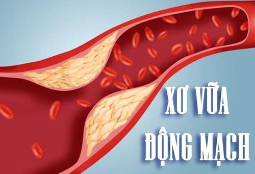
Xơ vữa động mạch là dấu hiệu cảnh báo phình động mạch chủ
● Accurately locate the abnormal aorta.
● Determine the maximum diameter at which the aortic segment is dilated, will be measured based on the diameter of the outer wall of the aorta and placed perpendicular to the axis of blood flow. At the same time, measure the length of the abnormal aortic segment.
● For patients with suspected or confirmed genetic syndrome at high risk of aortic disease, measurement of the annulus size and diameter of the sinuses of Valsalva is recommended. along with the diameter of the junction of the sinus of Valsalva and the ascending aorta, the diameter of the ascending aorta.
● Assess for the presence of atherosclerotic or thrombotic lesions on the surface of the vascular endothelium.
● Further evaluation for the presence of hematoma lesions appearing in the vessel wall, or atherosclerotic ulcers and calcifications.
● Determine the extent of aortic lesions with arterial branches including aneurysms and dissections, and evidence of secondary target organ damage such as decreased intestinal or renal perfusion.
● If previous imaging studies are available, direct comparisons can be made between images to detect an increase in the size of blood vessels.
4. Treatment
4.1. Medical
When suspecting that a patient has aortic dissection, emergency blood pressure lowering is required even though a definitive diagnosis has not been received:● Criteria: lower the patient's blood pressure to 100-120 mmHg and decrease the circulating blood flow. through the aortic valve. At the same time, it is necessary to try to stabilize the blood pressure value and try to reduce the blood pressure as much as possible and reduce the heart rate.
● Drugs to choose: Labetalol is a beta and alpha blocker that works to reduce the pressure of the vessel walls and control blood pressure quickly.
In addition, pain relief is also a way to better control blood pressure. Doctors can add Morphine Sulphate into the body intravenously, need to accurately detect the appropriate dose for the patient, for example: Morphine 10 mg needs to be diluted to 10ml with 0.9% sodium chloride, and then Intravenous injection of 5 mg, after 3-5 minutes, re-evaluate the pain situation, if not, then inject 5 mg again.
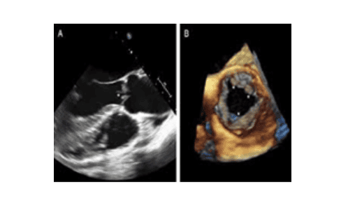
Siêu âm 4D van động mạch chủ
4.2. Surgery
In many cases of Type A aortic dissection, the patient will be indicated for surgery, the patient needs to be operated at a reputable unit, with a surgical team and professional anesthesia and resuscitation. Surgery can help to connect or replace the segment, or replace the aortic valve if there is damage to the valves of the aorta and coronary arteries.For cases of type B aortic dissection aneurysm, if there are complications with limb ischemia or renal artery, or mesenteric artery,... then the patient may be indicated for surgery, but need endovascular intervention to modify, this is the most optimal option today.
Aortic dissection has many dangerous complications, which can threaten the life of the patient if not diagnosed and treated promptly, but it is possible to use imaging to determine the course of the disease. disease, without causing trauma and with many different methods, supporting the most accurate diagnosis of the condition of the disease. Immediately go to reputable medical facilities for the best diagnosis and treatment.
Vinmec International General Hospital is the address for examination, prevention and treatment of many respiratory diseases, including chronic obstructive pulmonary disease. The examination, diagnosis and treatment of diseases are carried out by qualified and well-trained doctors along with modern medical equipment, which will bring optimal treatment results to patients.
Master. Dr. Le Hong Chien has many years of experience working in the field of diagnostic imaging and interventional radiology (endovascular intervention and extravascular intervention). Before being a Doctor of Diagnostic Imaging - Intervention at Vinmec Times City International Hospital, Dr. Chien was a Doctor at the Department of Diagnostic Imaging, Hospital 19.8 - Ministry of Public Security and Hong Ngoc General Hospital.
Customers can directly go to Vinmec Health system nationwide to visit or contact the hotline here for support.


