This is an automatically translated article.
This article is professionally consulted by Master, Doctor Bui Ngoc Phuong Hoa - General Internal Medicine - Department of Medical Examination & Internal Medicine - Vinmec Danang International Hospital.Voltage fluctuations are recorded as waves and have different manifestations in different neurological diseases. Based on the results of the EEG, the doctor will have a treatment plan suitable for each type of disease.
1. What is an EEG?
Using electrodes placed on the scalp, an electroencephalogram (EEG) is a recording of the bioelectrical activity of the brain. Metal electrodes attached to the skin on the outside of the head change electrical activity into patterns, called brain waves. The brain waves are recorded by the polyphone. A baseline EEG takes about 45 minutes, which can range from 30 minutes to 90 minutes.2. An overview of the EEG
EEG has 4 basic wave types: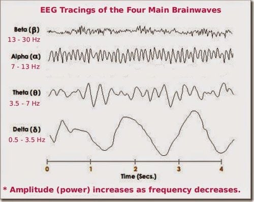
4 loại sóng não
Physiological disturbances: Electrocardiogram, electromyography, eye movements, tongue movements, respiration, skin perspiration,... Non-physiological noise waves: Due to environmental factors, due to device or possibly electrodes. Characteristics of a normal EEG:
For children: There is a lower frequency in the posterior dominant rhythm, a higher amplitude and sometimes a posterior slow wave. For adults: Posterior dominant rhythm at 8.5-11Hz, fast, low-frequency activities in the anterior leads. In some cases normal variation: slow alpha variation, fast alpha, lamda wave, Mu rhythm,... Some common abnormalities on EEG:
Abnormalities in amplitude: EEG isoelectric . Due to damage to the cerebral cortex, the lesion occupies the space inside or outside the skull, so the amplitude is low locally. In the case of patients with anxiety reducing the synchronicity of cortical activity leading to diffuse low amplitude, cortical damage or cortical dysfunction may also cause similar results. High amplitude beta activity, increased sharpness of the waves leads to locally increased amplitude in the cranium. Slow waves: Structural damage to the brain, either related to epileptic activity, or possibly due to encephalopathy that causes slow waves to appear. Cyclic Activity: General Cyclic Activities: Occurrence in metabolic encephalopathy, hypoxia, poisoning, mad cow disease. Periodic activities are localized in one hemisphere, sometimes occurring independently on both hemispheres. Brain damage such as stroke, encephalitis or chronic cases of epilepsy can all catch this abnormality. For cases with severe encephalopathy, there are two types: burst - suppression pattern or suppression - burst pattern. Seizures: The EEG will show whether there are epileptic discharges, general or local, and where they are located. Sharp spike, spike - slow wave complex, spike - wave complex, multi spike, multi spike - slow wave, multi spike - wave, multi spike - slow wave.
3. Benefits of EEG
EEG method helps patients find abnormal electrical brain waves in some neurological diseases. Brain wave patterns will be examined and monitored for abnormal wave patterns that lead to seizures and other problems. From the electrical brain waves during the EEG measurement, it is possible to diagnose headaches, epilepsy, ..., thereby giving appropriate treatment.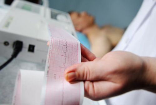
Lợi ích đo điện não đồ
Check, diagnose and monitor epilepsy or seizure disorder. Help support the diagnosis of brain death. Assess level of anesthesia. Besides, doctors can also diagnose the following diseases:
Sleep disorders. Dementia. There are brain tumors. Have encephalitis. Brain dysfunction. Stroke.
4. Harm of EEG
When the electrodes are attached to the scalp, the patient will feel some discomfort. However, EEG is a safe and harmless method, without any electrical current being passed into the body, it only records the electrical activity of the brain.5. Before measuring EEG, what should be prepared?
Clean the tip of the hair before the day of the EEG measurement. Do not drink coffee on the day of the EEG. If you are taking medication or have a pacemaker, tell your doctor. Patients must stay up late and get up early if sleep EEG measurements are required. While waiting for EEG measurement, absolutely do not sleep. Follow your doctor's advice exactly. Currently, Vinmec Hai Phong International General Hospital is a prestigious address in the examination and treatment of neurological diseases in general and headaches in particular:The hospital has modern equipment and machinery: The first hospital in Hai Phong to own a Signa Pioneer 3.0 Tesla Magnetic Resonator from GE (USA) and a CT scanner Aquilion One (Vision Edition) 640 slices from Toshiba (Japan), early and accurate diagnostic tools. damage to blood vessels and nerves. Currently, only Vinmec hospital in Hai Phong has a video EEG machine, which is of high value in accurately diagnosing epilepsy. In addition, other testing systems of the hospital are modern and ensure the correct process. A team of highly specialized medical doctors, examined by Specialist Doctor II. Pham Thi Son - Department of Medical Examination & Internal Medicine, Vinmec Hai Phong International General Hospital has 20 years of experience in the field of neurology. An EEG is one of several tests that can be used to find the cause and diagnose a headache. However, not all patients with headaches need this test. Only when prescribed by specialized doctors, patients can be performed.
Please dial HOTLINE for more information or register for an appointment HERE. Download MyVinmec app to make appointments faster and to manage your bookings easily.