This is an automatically translated article.
The article was professionally consulted with a Radiologist, Department of Diagnostic Imaging - Vinmec Central Park International General Hospital.Indications for digitizing spinal arteriography with background erasure are indicated by doctors when patients with arteriovenous malformations are at high risk of marrow rupture. The following article will help you better understand the process of digitizing spinal arteriography to erase the background.
1. What is a spinal artery?
The spinal cord is located inside the spinal canal, surrounded by three membranes: the outer membrane is the dura mater, the middle membrane is the arachnoid membrane, and the inner membrane is the feeding membrane. The spinal cord runs along the inside of the spine, in the spinal cord contains nerves that make connections from the brain to the whole body.In the spinal cord consists of gray matter in the middle and surrounded by white matter. The spinal cord has 3 longitudinal arteries, of which 1 is anterior and 2 is posterior. The three spinal arteries run vertically from the cervical medulla to the apex. The spinal artery forms from the root artery.
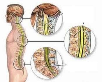
Tủy sống nằm xuyên bên trong ống cột sống, được bao bọc ba lớp màng
2. What is background digitization angiography?
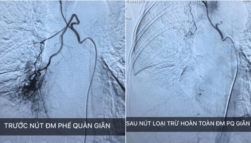
Digital erasure angiography is an iodinated contrast-enhanced angiogram to visualize the arteries supplying the spinal cord from the neck to the apex. Diseases involving the spinal cord may supply blood from the spinal artery or from the intercostal branches or from the carotid artery.2.1 When is digitized spinal angiography indicated?
When the patient has a high-risk arteriovenous malformation or ruptured marrowThe doctor needs embolization to reduce the size of the malformation and reduce the risk.
2.2 Process of digitizing spinal arterioles with background erasure
2.2.1 Preparation
To perform the technique of digitizing spinal arteriography to erase the background, it is necessary to have 1 specialist doctor, 1 nurse, and 1 electro-optical technician. The patient was thoroughly explained about the procedure to coordinate with the doctor during the procedure. Fasting, drinking before 6 hours. You can drink water but not more than 50ml of water. Usually digitized spinal angiography requires only local anesthesia. But for cases of children under 5 years old, or those who cannot coordinate with doctors, anesthesia will be indicated.2.2.2 Means and machines serving the indications for background erasure digital angiography
Digital erasure angiography machine (DSA) Specialized electric pump Film, film printer, image storage system Lead jacket, apron, X-ray shielding. General anesthesia, local anesthesia. The patient lies supine on the scanning table, intravenous line is placed (usually 0.9 % isotonic saline serum).
Gây mê toàn thân được chỉ định cho những bệnh nhân không phối hợp được với bác sĩ
2.2.3 Choosing the technique of use and the inlet of the catheter
Using the Seldinger technique, the catheter access route can be: From the femoral artery, the axillary artery, the brachial artery, the primary carotid artery, and the radial artery. Usually most are from the femoral artery, unless this is not possible, other access routes are used. Background digitized angiography. Sterilize and anesthetize the puncture site. Insert the needle and insert the tube into the lumen. Neck area: Selective catheterization of the vertebral arteries on both sides, body and shoulder, and then inject drugs. The dorsal and apical spinal cord: Insert a 4 -5F catheter into each intercostal artery for injector imaging. Look for the Adamkiewicz artery, usually at the level of the T7-T9 thoracic vertebrae. Inject the contrast agent through the machine with a volume of 6ml, speed of 2ml/s, pressure of 300 PSI. Recording and taking series of films focusing on the skull in an upright position, can take more inclined or oblique films when there is an injury. After satisfactory imaging, the catheter and catheter were removed and applied manual pressure directly on the puncture site for about 15 minutes to stop bleeding, then applied pressure for 8 hours. Any questions that need to be answered by a specialist doctor as well as if you want to be examined and treated at Vinmec International General Hospital, you can contact Vinmec Health System nationwide or register online. line.Please dial HOTLINE for more information or register for an appointment HERE. Download MyVinmec app to make appointments faster and to manage your bookings easily.


