This is an automatically translated article.
Diagnosing meningeal hernia in newborns plays an extremely important role in helping to detect the disease and treat it promptly, limiting the dangerous complications that the disease causes in children.1. What is meningeal hernia?
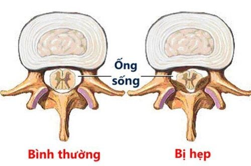
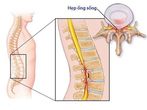
Meningeal herniation is a rare disease that occurs due to birth defects in the spinal canal (the hollow space of the vertebrae, which contains the spinal cord and nerve roots) of a child. Specifically, because the posterior arch of the vertebrae is wide open, the spinal canal communicates with the soft outside of the spinal canal, making the dura mater easy to bulge and form a hernia sac.
Meningeal herniation in infants is a serious medical condition, causing loss or dysfunction of nerve function, affecting shape and activities.
People divide meningeal hernia into the following types:
Meningocele: Hernia sac contains dura, arachnoid, and cerebrospinal fluid. Meningomyelocele: The herniated sac contains the dura mater, cerebrospinal fluid, and part of the spinal cord (or ponytail). Myelocele: The hernia sac fills the marrow. Syringomyelocele: Hernia sac contains the pulp and central canal.

Thoát vị màng não tủy là một tình trạng bệnh lý nặng thường gặp ở trẻ sơ sinh
2. Diagnosis of meningeal hernia in infants
Conspicuous feature of meningeal hernia in children is the appearance of tumors in the lumbar - sacral region. This tumor is usually soft, covered with wrinkled skin, inside is cerebrospinal fluid or a mixture of brain tissue and cerebrospinal fluid.
In terms of morphological characteristics, the hernia mass can have different shapes as follows: the skin is relatively thick, rarely breaks, causing cerebrospinal fluid leakage, the skin is thin, stretchy, easy to tear, causing cerebrospinal fluid leakage. marrow. The skin and subcutaneous fat layer in the hernia sac is quite thick, palpating externally like a tumor.
Some other features of the hernia sac are often described as concave when pressed but then rapidly swell. This hernia sac can change size according to the heart rate and breathing rate of the child.
Meningeal hernias usually do not show any motor and sensory disturbances. However, the disease can cause partial or complete paralysis of the legs, loss of sensation, and sphincter disturbances common in cases of hernia sacs with nerve roots and spinal cord (can be detected when shine a flashlight through the hernia sac).
Patients often have malformations in the high cervical spine and posterior cranial fossa called Anold - Chiari syndrome, which is a congenital malformation of the occipital region, causing the medulla oblongata and two cerebellar amygds to descend into the Cl and C2 spinal canals. Squeezing Sylvius and foramen of Magendie cerebrospinal fluid does not enter the subarachnoid space, causing hydrocephalus.
Diagnosis of meningeal hernia through the following methods:
Lumbar puncture shows the circulation of cerebrospinal fluid is obstructed. X-ray of the spine will show the position and extent of the posterior arch defect, and spina bifida in most cases. CT-Scanner: It will also determine the position and degree of defect of the posterior arch of the spine, but cannot evaluate the soft tissues, ligaments or meninges. Magnetic resonance imaging (MRI) of the lumbar spine: MRI is currently the most valuable imaging test for diagnosing myelomeningocele.
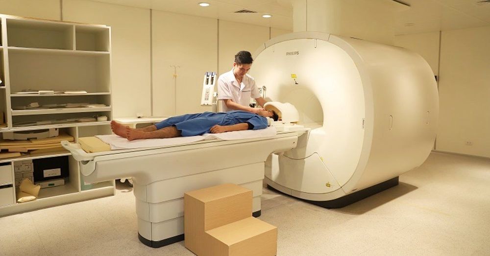
Hệ thống máy chụp cộng hưởng từ MRI tại Bệnh viện Đa khoa Quốc tế Vinmec giúp chẩn đoán bệnh lý thoát vị tủy– màng tủy chính xác
3. How to treat meningeal hernia?
Surgery is the optimal method to treat meningeal hernia. If detected early, the baby can be operated within the first 24 hours after birth. However, surgery for children with meningeal hernia is extremely complicated. Children need to be prepared very carefully before surgery, assess the degree of nerve damage. After surgery, children need to be closely monitored for complications, especially infection.
Please dial HOTLINE for more information or register for an appointment HERE. Download MyVinmec app to make appointments faster and to manage your bookings easily.


