This is an automatically translated article.
The article was professionally consulted by Specialist Doctor I Nguyen Truong Duc - Radiographer - Department of Diagnostic Imaging and Nuclear Medicine - Vinmec Times City International General Hospital.Computerized tomography of the lumbar spine is a technique that helps doctors check and evaluate pathologies in this area such as spinal malformations, spinal stenosis, herniated discs, etc. provide the most appropriate treatment for the patient.
1. An overview of the lumbar spine
1.1 Anatomical features of the lumbar spine
The lumbar spine is the central structure of the body, supporting the entire body. The lumbar spine consists of 5 vertebrae (symbols from L1 - L5), located between the rib cage and the pelvis, with the characteristics of a large vertebral body, wide in width; peduncle short, large diameter; transverse process thin, narrow and long; the spine is transversely directed posteriorly; The superior joint process is flattened horizontally, the inferior joint process is convex cylindrical.The lumbar spine consists of 7 parts: vertebral joints, intervertebral discs, lumbar graft, lumbar spinal canal, lumbar ligaments, spinal nerves and roots, vertebral motor segments. Large blood vessels, nerves, ligaments, tendons and cartilage are also part of the lumbar spine.
1.2 Functions of the lumbar spine
● Helps the body to move flexibly, bend, bend easily;● Protect the spinal canal, allowing nerve roots to govern the body's activities;
● Linked with the ribs into a solid framework for the muscles to attach to, protecting the body's internal organs.

Cột sống thắt lưng giúp cơ thể vận động linh hoạt, dễ dàng
1.3 Common diseases of the lumbar spine
Low back pain is a common problem in many age groups today. The main causes of back pain include: herniated discs, spine spurs, spondylolisthesis, arthritis and cancer. To avoid the risk of diseases in the lumbar spine, each person needs to create a healthy lifestyle and have regular health checkups. Computed tomography is a widely used technique to detect and evaluate pathologies in the lumbar spine.2. Learn about lumbar spine computed tomography
2.1 What is computed tomography of the lumbar spine?
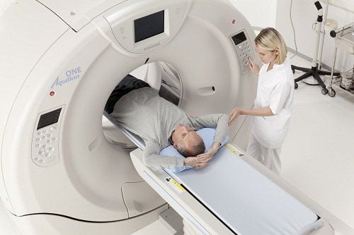
Computerized tomography (also known as computed tomography - CT scan) is a tomography x-ray technique, the data obtained will be reproduced by the computer system to create cross-sectional images of a certain part. in the body. When done, the machine will scan around the patient's body, send the acquired images to the computer and those images will be synthesized and analyzed by the electro-optical technician. In patients with a CT scan of the lumbar spine, the doctor will take cross-sectional images of the patient's back.2.2 Indications for performing computed tomography of the lumbar spine
CT scans produce detailed images of the waist quickly. This imaging technique can be used to check for the following:Birth defects of the spine;
Herniated disc;
Spinal stenosis ;
● Injury to the lower spine.
● Orientation for spinal intervention and post-treatment evaluation.
● Evaluate spinal problems in patients with contraindications to MRI.
A CT scan of the lumbar spine may also be used during or after an X-ray of the spine and spinal cord or an X-ray disc imaging (optical discography).

Bệnh nhân thoát vị đĩa đệm được chỉ định chụp cắt lớp vi tính cột sống thắt lưng
2.3 Contraindications
Computed tomography of the lumbar spine has no absolute contraindications. This technique is relatively contraindicated for pregnant women.3. The procedure of computerized tomography of the lumbar spine without contrast injection
3.1 Before CT
● Patient wears loose, comfortable clothing;● The patient is instructed to remove all metal objects on the body. The patient should tell the doctor if there are any metal fragments in the body from previous procedures;
● The patient tells the doctor all his health problems.
3.2 Implementation process
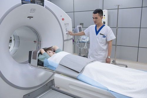
● The patient lies on a narrow table that can slide into the center of the CT scanner;● When the patient is inside the scanner, the machine will emit X-ray beams around;
● The computer picks up the signal and converts it into separate images of the spine region, called slices. Images can be stored, viewed on a computer screen, or printed into a movie. It is possible to combine slices together to create a 3-D image of the lumbar spine.

Hình ảnh chụp cắt lớp vi tính được xử lý qua phần mềm
3.3 After computed tomography of the lumbar spine
● Patient wears normal clothes and activities;● The images obtained after the CT scan will be processed and reproduced by the radiologist, the radiologist will read and return the results, advise the patient or request a CT scan again. , blood tests or other tests to confirm the diagnosis, and to choose the appropriate treatment if there is disease in the lumbar spine.
3.4 Read the results of computed tomography of the lumbar spine
● Normal results: If no problems are detected on the CT scan of the lumbar region;● Abnormal results: Warning of health problems such as birth defects of the spine, fractures, stenosis of the lumbar spine, herniated discs, spondylolisthesis,...
Note:
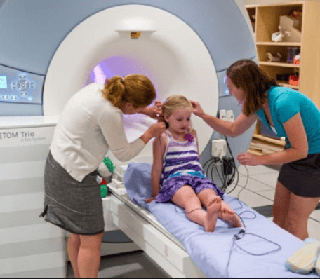
● Children Children need to follow the doctor's special instructions when having a CT scan. For children who are too young, cannot sit still, they should use sedatives;
● During the CT scan, the patient must lie still because moving can cause the image to be blurred. The person may also be told to hold their breath for short periods of time;
● After the scan, the patient may be asked to wait while the medical staff recheck the images to ensure that the patient's health problem can be accurately diagnosed;
● The CT scan only takes about 10-15 minutes.

Khi chụp CT cho trẻ em cần chỉ định tiêm thuốc an thần để tránh kích động
Vinmec International General Hospital is a high-quality medical facility in Vietnam with a team of highly qualified medical professionals, well-trained, domestic and foreign, and experienced.
A system of modern and advanced medical equipment, possessing many of the best machines in the world, helping to detect many difficult and dangerous diseases in a short time, supporting the diagnosis and treatment of doctors the most effective. The hospital space is designed according to 5-star hotel standards, giving patients comfort, friendliness and peace of mind.
Doctor Duc has nearly 17 years of experience in diagnostic imaging at Hanoi E hospital. Currently, the doctor is working at the Department of Diagnostic Imaging - Vinmec Times City International Hospital.
To register for an examination at Vinmec International General Hospital, you can contact the nationwide Vinmec Health System Hotline, or register online HERE.


