This is an automatically translated article.
The article is expertly consulted by Master, Doctor Hoang Van Lan Duc - Doctor of Radiology - Department of Diagnostic Imaging and Nuclear Medicine - Vinmec Times City International General Hospital.Computed tomography (CT) scan of the lumbar spine with 3D rendering is a technique performed to diagnose problems in the spine - lumbar of the patient. From there, the doctor can make the right treatment option for the patient.
1. Lumbar spine characteristics
The lumbar spine is the structure that supports the weight of the entire body, so it plays a very important role. According to a survey, up to 80% of the world's population at least once in their life has problems related to the lumbar spine.1.1 Anatomy
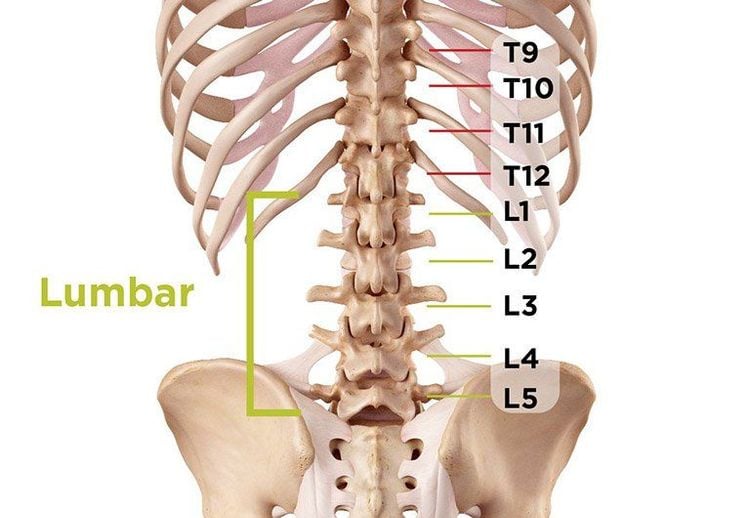
Anatomically, the lumbar spine has 5 vertebrae with symbols from L1 to L5. The lumbar spine is located between the rib cage and the pelvis. It has the following characteristics: The vertebral body is large, wide in width; short bow peduncle of large diameter; The transverse process is thin, narrow and long, the spine is transversely directed posteriorly.Internal structure of the lumbar spine consists of 7 parts:
Vertebral joint: Includes cartilage, synovial fluid, synovial capsule and joint capsule. Vertebral joints have limited mobility, only moving in the anterior and posterior directions, moving in the longitudinal direction of the body; Intervertebral disc: It is composed of cartilage plate, fibrous ring and mucus nucleus. Lumbar disc has a thickness of about 9mm, there is a difference in the thickness of anterior and posterior discs depending on the physiological curvature of the spine; Lumbar graft hole: The place where the nerves pass, made up of the inferior and superior defects of the upper and lower vertebrae. The graft holes are often pinched, causing low back pain;

Hình ảnh giải phẫu cột sống thắt lưng
1.2 Functions
The lumbar spine has a similar function to the other spinal segments. They are:Helps the body to move flexibly, easily bend, bend; Protecting the spinal canal: This is the starting point of the nerve roots, which plays a very important role in ensuring that the body's activities are governed; Linked with the ribs to form a solid frame, helping the muscles to attach and protect the internal organs of the body.
1.3 Common diseases of the lumbar spine
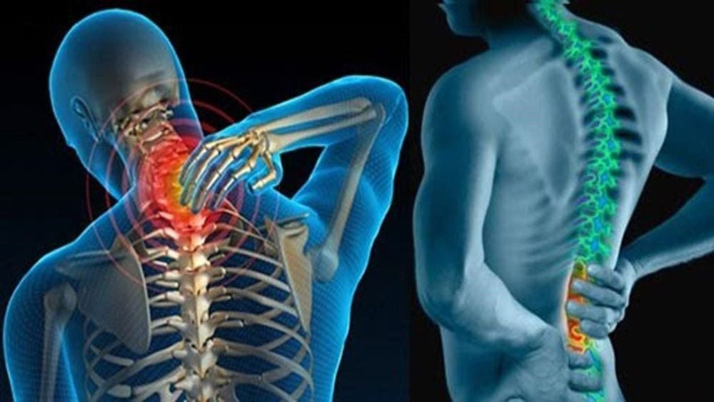
Office workers, people who are inactive or often work often have low back pain. Symptoms of pain are: Pain originating from the upper back; acute pain (days to weeks) or chronic pain (more than 3 months); pain may radiate along the spine down the leg; often have a lot of pain in the early morning or at night, making it difficult to sleep; pain that gets worse with prolonged sitting or movement; difficulty standing upright, limited mobility; can lead to fever, chills, and loss of bowel and bladder control.Low back pain can occur in all subjects, all ages. This symptom is a manifestation of a number of diseases such as: Spinal spine, herniated disc, spinal degeneration, arthritis, cancer,... CT scan of the lumbar spine with 3D rendering is a technique. effective diagnosis of this condition.

Gai cột sống là bệnh lý thường gặp về cột sống thắt lưng
2. The process of taking CT scan of the lumbar spine with 3D rendering
This is a technique that uses a computer tomography machine to create images of the lumbar spine, helping to evaluate the damage of bones, spinal canal and adjacent components. In principle, a body organ through computerized tomography will produce an infinite number of slices, if they overlap, it will give the original shape. The 3D rendering mechanism is a sequential rendering of multiple slices, layer by layer, until the complete form of the organ to be diagnosed (in this case, the lumbar spine).2.1 Indications and contraindications
Indicated in the diagnosis and evaluation of traumatic, tumor, and inflammatory diseases of the bones and soft parts of the lumbar spine; Contraindications: This technique has no absolute contraindications but relative contraindications for pregnant women.2.2 Preparation for implementation
Implementation personnel: Electro-optical technicians, specialists and nurses; Technical facilities: Including multi-slice computerized tomography machine (over 8 rows), film, film printer and image storage system; Patient: The doctor explains the technique to coordinate according to the instructions; remove metal body jewelry such as hairpins, necklaces, earrings; give sedation if the patient is too excited, can't lie still; Test sheet: There is a written order for the computerized tomography scan and relevant papers if any.
Bác sĩ có thể chỉ định người bệnh dùng thuốc an thần để tránh kích động
2.3 Technical implementation
Patient position: Put the patient in the machine frame, lie on his back, 2 loans lowered to the maximum, 2 hands raised up along the body axis. Patients need to hold their breath, do not swallow during the CT scan; Localization of the entire lumbar spine in 2 planes; Take the locator image in a lateral direction starting from the superior border of vertebra D12 to the inferior border of vertebra S1; Set the imaging program according to clinical requirements. Can use cutting layers in the direction of discs to evaluate pathology in the lumbar spine and use software that allows image processing after taking. Commonly used image processing techniques include: volumetric surface rendering (VRT), surface rendering (SSD), multi-sectional imaging (MPR) and dark intensity projection techniques. multi (MIP); Select film images on disc windows and bone windows.2.4 Evaluation of results
Creating 3D images in CT scan of the lumbar spine has special value for the change of lumbar spine posture. The reconstructed images in the frontal plane, the mid-sagittal plane are very meaningful. This method is used to:Evaluate the lesions encountered in degenerative spondylosis including spinal stenosis, spondylolisthesis due to degeneration, ligament degeneration and lateral joint block degeneration; Assess for signs of disc herniation, vertebral body injuries (sliding vertebrae, vertebral body rupture and vertebral collapse), displacement of posterior vertebral wall lesions, traumatic hematoma, vertebral soft tissue injury , spinal stenosis ; Assessment of congenital abnormalities in the spine; Consider anatomical correlation, degree of displacement, invasion and compression. The method of taking CT scan of the lumbar spine with 3D rendering did not have any complications. However, the patient may have to repeat the technique if the image is not clearly revealed, does not remain motionless during the imaging process,...

Thông qua ảnh 3D cho phép bác sĩ chẩn đoán hình ảnh về tổn thương bệnh lý thoái hóa đốt sống
Vinmec International General Hospital is a high-quality medical facility in Vietnam with a team of highly qualified medical professionals, well-trained, domestic and foreign, and experienced.
A system of modern and advanced medical equipment, possessing many of the best machines in the world, helping to detect many difficult and dangerous diseases in a short time, supporting the diagnosis and treatment of doctors the most effective. The hospital space is designed according to 5-star hotel standards, giving patients comfort, friendliness and peace of mind.
Master. Doctor Hoang Van Lan Duc was formally trained and graduated with a Master's degree from Hanoi Medical University. The doctor has more than 10 years of experience in the field of imaging, especially in diagnosing with emergency internal, surgical, abdominal, thoracic, musculoskeletal, neurological and thyroid, breast, ...
To register for an examination at Vinmec International General Hospital, you can contact the nationwide Vinmec Health System Hotline, or register online HERE.


