This is an automatically translated article.
The article is expertly consulted by Master, Doctor Trinh Thi Phuong Nga - Radiologist - Department of Diagnostic Imaging and Nuclear Medicine - Vinmec Times City International General Hospital.Abdominal computed tomography and subframe computed tomography is a technique that uses a tomography machine to reconstruct images of the abdominal and pelvic structures to diagnose and look for pathological abnormalities, if any. The non-contrast abdominal/subframe computed tomography procedure is simple and non-invasive, and the risk of radiation exposure is very low.
1. What is a non-contrast computed tomography of the abdomen/subframe?
Computerized tomography of the abdomen/subframe without contrast injection is a technique that uses a multi-sequence computerized tomography machine, applying X-rays and algorithms to capture, process, and reconstruct all structural images. Abdominal, pelvic region accurately and in detail, thereby, allowing detection of lesions, abnormalities, pathologies in the abdomen and pelvis such as infection, trauma, appendicitis, kidney stone disease , gallstones, cysts, or tumors.Currently, the commonly used abdominal/subframe computed tomography machine is a multi-sequencing system (2, 16, 32, 128 rows, ...), which can capture the entire abdominal system. , subframe from the diaphragmatic arch to the pubic joint.

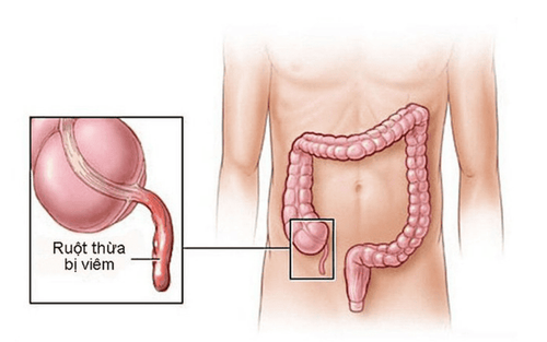
Chụp cắt lớp vi tính bụng/tiểu khung không tiêm thuốc cản quang giúp phát hiện bệnh viêm ruột thừa
2. Indications for computed tomography of the abdomen/subframe without contrast injection
Computed tomography of the abdomen/subframe without contrast is indicated to detect lesions in the following organs:● Liver: Liver injury, inflammation, cyst, liver abscess, liver tumor.
● Gallbladder, biliary tract: Gallstones, tumors in the biliary tract or gallbladder.
● Pancreas: Pancreatitis, pancreatic tumor, pancreatic stones.
● Spleen: Spleen injury, spleen tumor.
● Two kidneys: tumor, inflammation, stone, fluid retention, deformity....
● Bilateral adrenal gland: tumor, bleeding....
● Bladder: tumor, stone ....
● Abnormalities in the pelvic region: tumor, abscess, bleeding....
● Gastrointestinal tract: Trauma, gastrointestinal bleeding, gastrointestinal inflammation, gastrointestinal tract tumor.
● Other injuries such as: abdominal abscess, pelvic cavity, mesenteric fatty mane, mesenteric tumor,...
● Injury to the retroperitoneal space: tumor, fibrositis, bleeding ....
● Abnormalities of the spine in the lumbar region, pelvis, and sacral region....
Note, it is important to consider the appointment of non-contrast computed tomography of the abdomen/subframe without contrast for pregnant women. the first weeks.

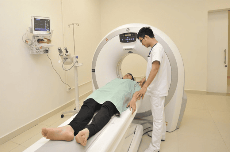
Quy trình chụp cắt lớp vi tính bụng/ tiểu khung không tiêm cản quang
3. Abdominal/subframe computed tomography without contrast injection
The procedure of computed tomography of the abdomen/subframe without contrast injection consists of the following steps:● Step 1: The patient is placed supine on the scanning table, arms raised above the head to limit interference. Patients are instructed and asked to hold their breath during the scan to avoid image noise.
● Step 2: The technician conducts tomography with a cross-section of the entire abdomen and subframe with the field from the diaphragm to the pubic joint. The thickness of the cut is in the range of 5-8 mm, with small lesions, the thickness of the cut is about 3 mm.
● Step 3: Depending on the size of the individual, the technician changes the field of view for the abdominal/subframe computed tomography scan accordingly. To evaluate the entire bone, fat, air, and soft tissues, the technician varies the width of the window.
● Step 4: The technician reproduces computed tomography images of the abdomen and subframe on the scanner at the imaging room. The resulting image must be clear, free from noise and show the entire structure of the captured area.
Computerized tomography of the abdomen/subframe without contrast injection is a modern, non-invasive imaging technique that allows the entire area to be evaluated to find, detect and determine the cause of the cause. abnormalities, if any.

Chụp cắt lớp vi tính bụng/tiểu khung không tiêm cản quang là kỹ thuật tiên tiến đã được áp dụng tại Bệnh viện Đa khoa Quốc tế Vinmec
Doctor Trinh Thi Phuong Nga is a radiologist with nearly 20 years of experience in diagnostic imaging. Currently, the doctor is working at - Department of Diagnostic Imaging - Vinmec Times City International General Hospital.
To register for examination and treatment at Vinmec International General Hospital, you can contact the nationwide Vinmec Health System Hotline, or register online HERE.


