This is an automatically translated article.
The article was professionally consulted by Specialist Doctor I Vo Cong Hien - Radiologist - Radiology Department - Vinmec Nha Trang International General Hospital.Hepatic artery chemoembolization is a modern, minimally invasive treatment for liver cancer that helps prolong and improve patients' quality of life. Computed tomography can be performed in various stages of the chemo node technique, making the technique more convenient and accurate.
1. Hepatic artery chemoembolization method in the treatment of liver cancer
Hepatic artery chemoembolization is a treatment for liver cancer that relies on the principle of blocking the blood supply to the tumor. The basis of this method is that the healthy liver parenchyma is supplied with 80% of blood from the portal vein system, 20% from the hepatic arteries. Meanwhile, the liver cancer mass receives 80% of its blood supply from the arterial system including the intrahepatic arteries and the extrahepatic arteries. Therefore, when injecting embolization materials and anti-cancer chemicals into the arteries feeding the tumor, the tumor will be ischemic, leading to necrosis.Liver artery chemotherapy is indicated in the following cases:
Liver cancer no longer requires surgery or surgery is possible but the patient does not want to. Ruptured hepatocellular carcinoma: many cases of ruptured cancerous nuclei have been successfully treated with hepatic artery chemoembolization. Vascular secondary liver cancer. There was no portal vein thrombosis, no large lymph node metastasis to the hilar liver. Hepatic artery chemoembolization is a modern treatment method with many advantages such as less invasiveness, short implementation time, high safety, few complications, few side effects, short hospital stay, .. This is an effective treatment for liver tumors, helping to prolong the patient's survival time and quality of life.
In addition, when the patient is found to have the following tumors, chemotherapy can be treated including:
Hepatocellular carcinoma (HCC) Cholangiocarcinoma (cell cancer of the pancreas) bile in the liver) Liver metastases include: colon cancer, breast cancer, neuroendocrine cancer. Large hemangiomas.
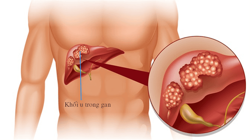
Bệnh nhân ung thư gan được chỉ định thực hiện
2. The role of computed tomography in hepatic artery chemotherapy
To perform hepatic artery chemoembolization, the support of computed tomography is extremely necessary. Computed tomography scan will help determine the angiogenesis in the liver tumor and locate the vascular peduncles feeding the liver tumor. When these important locations are identified, it will be more convenient and accurate to affect the artery feeding the tumor. In addition, computed tomography also helps to evaluate the results of embolization before the end of the intervention process.Currently, new generations of DSA angiograms can be used to perform computed tomography right on the angiographic table, this method is called cone-beam computed tomography (CBCT). CBCT is indicated in hepatic artery chemoembolization when the digitized background scan (DSA) cannot find a feeding artery, or the resulting image is not clear.
Computed tomography in the hepatic artery chemotherapy node is relatively contraindicated when the patient is not holding their breath or is not cooperating well. Patients with coagulopathy and severe cirrhosis need to be adjusted before intervention.

Máy chụp DSA cho phép chẩn đoán hình ảnh chính xác
3. Computed tomography procedure in the hepatic artery chemotherapy node
3.1. Prepare
The implementation team includes specialists, assistant doctors, radiology technicians, nurses and doctors, anesthesiologists (if the patient cannot cooperate). The means used to perform include: new generation DSA background eraser digital angiogram with CBCT scanning function; specialized electric pump; film, film printers, photo storage systems; lead vest, apron, X-ray shielding; necessary medicines and medical supplies.On the patient's side, the medical staff will explain the procedure carefully to coordinate with the doctor. Patients need to fast for 6 hours before the procedure, can drink water but not more than 50ml.
3.2. Steps to take
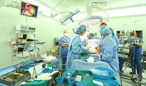
The patient lies supine on the intervention table. The team installed a machine to monitor breathing, pulse, blood pressure, electrocardiogram, SpO2. Sedative medication may be prescribed if the patient is restless and hyperactive. Then, disinfect the skin and cover with a sterile towel with holes.To enter the artery to be affected, access is usually from the right femoral artery, in special cases the brachial artery may be used.
Chemical hepatic embolization was performed by angiography of the visceral body, superior mesenteric vessels, and some suspected extrahepatic circulation. Then, a small ultra-selective catheter is inserted into the artery feeding the tumor and plugging it with embolic materials. Cone-beam computed tomography technique can be applied in many different stages in the intervention process such as:
To find the artery feeding the tumor: proceed to hook the tip of the 4-5F catheter in the trunk artery viscera or superior mesentery. Perform cone-beam computed tomography with a contrast pump, the dose of contrast injection is 5-6ml in 6 seconds. Do not perform imaging immediately after injecting the drug, but wait about 4-10 seconds for the contrast agent from the electric syringe to enter the liver vessels. Perfusion scan of liver parenchyma containing tumor: in cases where liver tumor does not really proliferate, digital erasure scan (DSA) will be very difficult to detect whether the selected blood vessel is really supplying blood to the tumor. u or not. To confirm, place the tip of the catheter in the suspected artery, then do a CBCT. CBCT scan before the end of the hepatic artery chemoembolization procedure: cone-beam computed tomography without contrast helps to assess whether the concentration of Lipiodol in the tumor is good. After the intervention, the patient lies in bed, immobilizes the punctured leg for at least 6 hours, and closely monitors the bleeding at the puncture site, pulse, blood pressure, and temperature. The patient will be given pain medication, fluids, and prophylactic antibiotics to fight infection. If the patient has a fever, a blood count and CRP will be done to rule out superinfection.
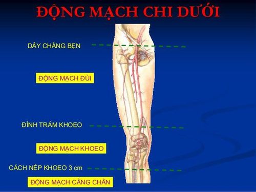
Đường vào động mạch trong nút mạch gan hóa chất thường là từ động mạch đùi bên phải
4. Possible complications
Bleeding at the puncture site, dissection of the abdominal aorta or the visceral-hepatic artery are complications associated with vascular intervention. The patient will be treated medically and closely monitored. If the condition is severe, the doctor may recommend surgery to treat. Hepatic embolism syndrome includes symptoms of pain, fever, elevated liver enzymes,... patients usually recover after 1 week. Inflammation and necrosis of the gallbladder: the patient will be treated medically, if it is not effective, the doctor may appoint surgery. Tumor necrosis causing liver abscess: usually requires percutaneous drainage and antibiotics. To perform hepatic artery chemoembolization, the support of computed tomography is extremely necessary. Computed tomography will help identify angiogenesis in liver tumors. However, to get accurate results, patients need to choose a reputable medical examination address with a full system of medical machines to perform.In order to meet the needs of medical examination and treatment, currently Vinmec International General Hospital has been and continues to bring in a system of modern machines such as magnetic resonance imaging (MRI), computed tomography (CT), X-ray, etc. modern DSA angiography room, ... in the work of medical examination and treatment, diagnostic imaging, disease treatment. With a full range of modern medical equipment, the implementation of the hepatic artery chemoembolization technique allows the doctor to control the entire procedure, avoiding maximum damage to the organs. organs in the body bring good quality of life to patients. In particular, at Vinmec, in addition to treatment with chemotherapy, the hepatic artery can also be combined with other treatment methods such as surgery, chemotherapy, and thermal tumor destruction (high-frequency ablation, microwave ablation, etc.) ...). For maximum efficiency.
At the present time, Vinmec also designs many accompanying medical services such as the package of screening and early detection of liver cancer suitable for many subjects at risk of different diseases, including the cancer screening package. liver - detect cancer early when there are no symptoms, contributing to early detection of liver cancer for timely treatment.
With a team of highly qualified medical professionals and modern machinery, the application of the TACE method at Vinmec International General Hospital has contributed to improving the quality of life. quality of life and improve patient survival.
BSCK I Vo Cong Hien has many years of experience in the field of imaging, especially in diagnostic ultrasound for general abdominal diseases, advanced cardiac - vascular, obstetrics and gynecology. Doctor Vo Cong Hien used to work at the Imaging Department of the University of Medicine and Pharmacy Hospital - Hoang Anh Gia Lai before working at the Imaging Department of Vinmec Nha Trang International General Hospital.
When customers have a need for medical examination and treatment at Vinmec, they can call the hotline or register for an online consultation with Vinmec HERE.