This is an automatically translated article.
This article is professionally consulted by Master, Doctor Vu Huy Hoang - Radiologist - Department of Diagnostic Imaging and Nuclear Medicine - Vinmec Times City International Hospital. Doctor Vu Huy Hoang has 10 years of experience working in the field of diagnostic imaging.Cone-beam computed tomography (CBCT) is a very effective technique in supporting radiofrequency ablation in the treatment of liver tumors. CBCT scans can be performed with new generation DSA angiograms.
1. What is cone-beam computed tomography in the treatment of liver tumors?
1.1. Radiofrequency ablation method to treat liver tumors
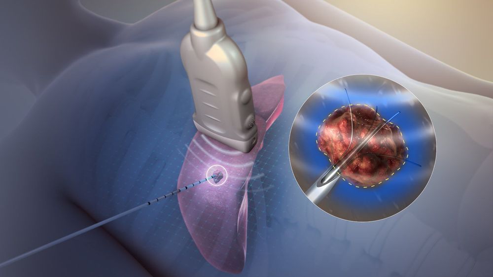
Radiofrequency ablation for liver tumors is an effective method to treat liver tumors less than 5cm in size. High-frequency ablation has particularly good results in patients with less than 3 tumors and each tumor size is less than 3cm. For larger liver tumors, radiofrequency ablation can be combined with other liver cancer treatments such as chemical embolization, absolute alcohol or acetic acid injections to increase the effectiveness of the treatment. result of high-frequency ablation.The principle of high frequency burning method is to convert alternating current into heat energy. A corrugated needle is inserted into the center of the tumor, an electric current is passed, generating heat to destroy the tumor. The electric current will be adjusted to ensure the generation of heat at 60-100 degrees Celsius. This is a temperature that can cause instant death of tumor cells. The time to perform radiofrequency ablation usually lasts from 8-12 minutes, depending on the specific case.

1.2. Cone-beam computed tomography in the treatment of liver tumors
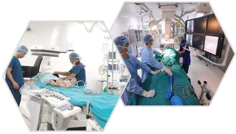
To ensure accuracy in the intervention process, the radiofrequency ablation method needs the support of imaging techniques. Usually, radiofrequency ablation is usually performed in the interventional room because these are rooms that are both as sterile as the operating room and supported by equipment such as DSA background angiography machine, ultrasound machine. sound, means of anesthesia resuscitation,..New generation DSA angiograms can be used for computed tomography. The method of computed tomography right on the angiogram table is called cone-beam computed tomography (CBCT).
Cone-beam computed tomography will be done at two times, that is:
Taken just before starting the radiofrequency ablation machine to make sure the ablation needle is in the correct position in the tumor need intervention. Taken after radiofrequency ablation to determine the extent of necrosis and residual tumor. The method is absolutely contraindicated in patients with coagulopathy. For patients with central liver tumors, subcortical liver tumors adjacent to the gastrointestinal tract structures, patients with a history of biliary-enteric anastomosis,... Doctors must consider very carefully before performing the procedure.

2. Preparation before performing cone-beam computed tomography in radiofrequency ablation for liver tumors
The implementation team includes a specialist, an assistant doctor, an radiologist, a nurse, a doctor or an anesthesiologist. The necessary equipment is a digital background eraser angiogram (DSA) with the function of cone-beam computed tomography (CBCT); specialized electric pump; high frequency wave therapy machine; film, film printer, image storage system with lead vest, apron to shield X-rays.On the patient's side, to prepare for the procedure, the patient will be consulted by medical staff about the condition of the patient. condition, carefully explain the procedure to coordinate with the implementation team. Patients need to fast for 6 hours before the procedure, can drink water but not more than 50ml.

3. Steps to perform cone-beam computed tomography in high-frequency ablation of liver tumors
In the intervention room, the patient lies supine on the operating table. The team installed a machine to monitor breathing, pulse, blood pressure, electrocardiogram, SpO2. Then, disinfect the skin and cover with a sterile towel with holes. If the patient is excited, not lying still, the doctor will prescribe a sedative. Patients receive pain relief by anesthesia, pre-anesthesia or general analgesia. The doctor inserts a high-frequency needle into the tumor through the skin under the guidance of ultrasound or computed tomography. Cone-beam computed tomography to make sure that the ablation needle is in the correct position at the center of the tumor. After you have made sure that the radiofrequency ablation needle is in the correct position, proceed to start the radiofrequency ablation machine. Depending on the size of the tumor, the treatment time can be from 8-12 minutes. Conduct ablation during the removal of the needle from the liver. Cone-beam computed tomography to determine the extent of necrosis and residual tumor. Radiofrequency ablation achieves good results when the area of the destructive wave needle is 1-1.5cm larger than the tumor area and does not cause damage to nearby organs.
4. Complications may be encountered when performing cone-beam computed tomography in radiofrequency ablation for liver tumors.
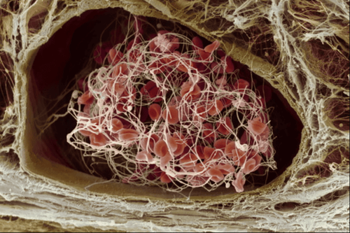
Common complications immediately after intervention are:Abdominal bleeding: The patient will be closely monitored for hemodynamic factors. If you are anemic, you will be given blood transfusions and clotting factors. If medical treatment is not effective, the doctor will recommend surgery to stop the bleeding. Hollow visceral perforation: Usually monitored medically and treated with antibiotics. If that doesn't work, the patient will be indicated for surgery to stitch the perforation. A common late complication is biliary atresia. If the patient is not clinically symptomatic, intervention may not be necessary. If the patient has complications of biliary tract infection, jaundice, surgery must be performed to dilate and drain the biliary tract.
In order to meet the needs of medical examination and treatment, currently Vinmec International General Hospital has been and continues to bring in a system of modern machines such as magnetic resonance imaging (MRI), computed tomography (CT), X-ray, etc. .. into the work of medical examination and treatment, diagnostic imaging, disease treatment. With a full range of modern medical equipment, the implementation of high-frequency ablation technique for liver tumor treatment allows the doctor to control the entire procedure, avoiding maximum damage. Injury to blood vessels, nerves, trachea, esophagus should be very safe, and at the same time reduce the maximum tumor size.
Especially to bring high efficiency in disease examination and treatment, Vinmec now also designs many accompanying medical services such as the package of screening and early detection of liver cancer suitable for many at-risk subjects. Risk of various diseases including the liver cancer screening package - detecting cancer early when there are no symptoms brings convenience and satisfaction to all customers.
Please dial HOTLINE for more information or register for an appointment HERE. Download MyVinmec app to make appointments faster and to manage your bookings easily.


