This is an automatically translated article.
The article was professionally consulted by Specialist Doctor II Tran Thi Mai Huong - Department of Obstetrics and Gynecology - Vinmec Hai Phong International General Hospital.1. The formation of the fetal heart
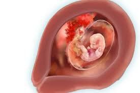
During pregnancy, on the 16th day of pregnancy, the embryo begins to develop two blood vessels that form the two ducts of the heart. Entering the 5th week of pregnancy, the fetus is 3 weeks old, the embryo begins to take shape and the neural tube has formed. At the end of the 5th week, the small seed in the middle of the embryo begins to form, making a premise for the development of the fetal heart later. However, the fetal heart only really grows when the fetus enters the 7th week of pregnancy.In the 7th week of pregnancy, the fetal heart grows and begins to divide into two left and right chambers. In the 11th week of pregnancy, the fetal heart begins to beat slightly and is almost complete until about the 12th week. In the 14th week, the fetal heart beats more clearly. By the 16th week of pregnancy, you can already pump blood with an amount of about 24 liters / day and this amount is likely to continue to increase with the growth of the baby. At this time, structurally the heart has begun to complete and take on its functions.
From the following weeks of pregnancy until the baby is born, the fetal heart will continue to grow, larger in size and volume. The normal heart rate is about 120-160 beats/minute.

2. When can the mother hear the baby's heartbeat?
In the 6th week of pregnancy, the baby's heart beats about 110 times / minute, and the heart rate increases to 150-170 beats / minute over the next 2 weeks, at this time, the baby's heart beats twice as fast as the mother's heart.According to this growth, the mother can hear the baby's heartbeat for the first time at the 9th or 10th week of pregnancy. The heart rate will be around 170 beats/minute and will slow down gradually. During the exam, if you want to listen to the fetal heart, the doctor will place a hand-held ultrasound device called a doppler on top of the mother's abdomen to amplify the sound.
In the 12th week of pregnancy, the baby's bone marrow begins to produce blood cells. Until week 17, the fetal brain begins to adjust the heart rate to prepare to support the baby in the outside world, at which point the heart is already beating naturally. Around the 20th week of pregnancy, you can hear your baby's heartbeat through a stethoscope.
3. When to ultrasound and evaluate for congenital heart defects
Fetal heart rate ranges from 120-160 beats/minute, but can increase to 180 times/minute if the baby is moving a lot in the womb. Fetal heart rate by week of pregnancy will also change. However, mothers should note that if the fetal heart rate is less than 120 beats / minute at 6-8 weeks, there is a very high risk of miscarriage.Cases of fetal heart rate below 110 beats / min are considered bradycardia. There are many causes of slow fetal heart rate such as poor blood circulation, low blood pressure in pregnant women, fetal malformations or placental anomalies.
From the 6th to the 9th week of pregnancy, the doctor will perform the first trimester ultrasound to confirm the pregnancy, calculate the gestational age and check the fetal heart is healthy. A fetal echocardiogram from 18 to 24 weeks will help the doctor evaluate more deeply. Especially for mothers with a family history of congenital heart defects, or phenylketonosis, diabetes or autoimmune disease, ... should inform the doctor.

In addition, in some cases, surgery may be necessary immediately after birth. Some other disabilities have to wait until the baby is older to be operated on, or treated with drugs.
4. Baby's heart at birth
The fetal circulatory system will continue to develop steadily until about 40 weeks, when it is ready for birth. The function of the circulatory system is very different when the baby is inside the uterus and at birth. Before birth, the baby's lungs are not functioning because the baby cannot breathe in the womb. The umbilical cord supplies oxygen- and nutrient-rich blood to the developing circulatory system.The fetal heart has two shunts, the foramen ovale and the ductus arteriosus. The foramen ovale connects the right atrium and the left atrium. Blood from the umbilical vein of the fetus flows to the inferior vena cava, to the right atrium, from here the blood passes through the foramen ovale located between the two atria to the left atrium, down the left ventricle, up the aorta to the nourishment. the upper organs of the fetus.
Then follow the superior vena cava back to the right atrium, down the right ventricle, into the main common artery. Because the lungs are not yet functioning, only 10% of the blood goes to the lungs to feed the lungs, and 90% through the ductus arteriosus into the descending aorta to nourish the lower organs of the fetus. Blood from the right atrium through the left atrium, from the pulmonary artery through the ductus arteriosus through the aorta is because the pulmonary vascular system pressure is higher than the aortic vascular system pressure.
When the baby is born, the baby's lungs begin to function, blood no longer goes from the pulmonary vascular system through the aortic vasculature, so the foramen ovale and ductus arteriosus gradually close to completely blocked, the circulatory system children act like normal people.

5. What should mothers do to help their children's hearts be healthy?
Pregnancy is a very sensitive time, the baby is constantly developing and there are many changes in the womb. The external environment can affect the development of the baby's cells. To help the baby's heart healthy and to help the development take place normally, pregnant women should note:Take folic acid and choose foods containing folic acid before and during pregnancy. This can prevent congenital heart disease in the baby. Do not use stimulants such as tobacco, alcohol,... Well control blood sugar during pregnancy if the mother has type 2 diabetes or gestational diabetes. Do not use drugs that are not recommended for pregnant women such as acne medicine Accutane,... The formation of a fetal heart is a sign of the existence of the fetus in the womb, a period of time. especially during pregnancy. At this time, mothers need to pay attention to the fetal heart rate, regular and scheduled fetal ultrasounds to check the cardiovascular status as well as the child's development, in order to detect birth defects early and have appropriate methods. early treatment.
Vinmec International General Hospital offers a Package Maternity Care Program for pregnant women right from the first months of pregnancy with a full range of antenatal care visits, periodical 3D and 4D ultrasounds and routine tests to ensure that the mother is healthy and the fetus is developing comprehensively.
Pregnant women will be consulted and checked for health under the close supervision of experienced and specialized Obstetricians, helping mothers have more knowledge to protect their health during pregnancy as well as reduce reduce complications for mother and child.
Doctor Tran Thi Mai Huong has 25 years of experience in examination and treatment in the field of Obstetrics and Gynecology, lower tract surgery, laparoscopic surgery. Has held the position of deputy head of the School of Obstetrics and Gynecology, deputy head of the delivery department - Hai Phong Obstetrics and Gynecology Hospital. Having high expertise and strength in the fields of:
Lower tract surgery Laparoscopic surgery Difficult obstetric surgery If you have a need for consultation and examination at Hospitals under the national health system, you are welcome. Please book an appointment on the website for the best service.
Please dial HOTLINE for more information or register for an appointment HERE. Download MyVinmec app to make appointments faster and to manage your bookings easily.


