This is an automatically translated article.
At some point in our lives, we are all overwhelmed by fear and hear many advices from friends like: “Think hard, listen to your brain, there's no excuse. To be scared!”. You should be able to explain to them that the epicenter of fear is in the brain itself, and it's called the amygdaloid in the body. In this article, we will focus on the anatomical structure of the amygdala, and the roles this structure plays in emotional response, decision-making, and memory.1. Anatomical structure of the amygdala
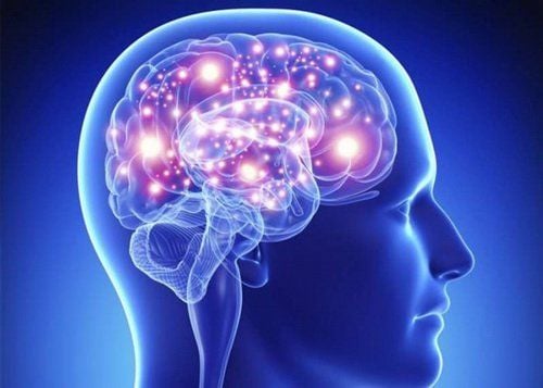
1.1. The Amygdala is one of two groups of almond-shaped nuclei located deep in the temporal lobe, medial to the hypothalamus and adjacent to the thalamus and inferior (temporal) horns of the lateral ventricles.In general, the main functions of the amygdala are strongly correlated with unpleasant and aversion situations and stimuli, and consequent responses to them. These types of stimuli have great biological significance for evolutionary reasons, and the amygdala is therefore one of the brain regions most important for such "primitive" behaviors.
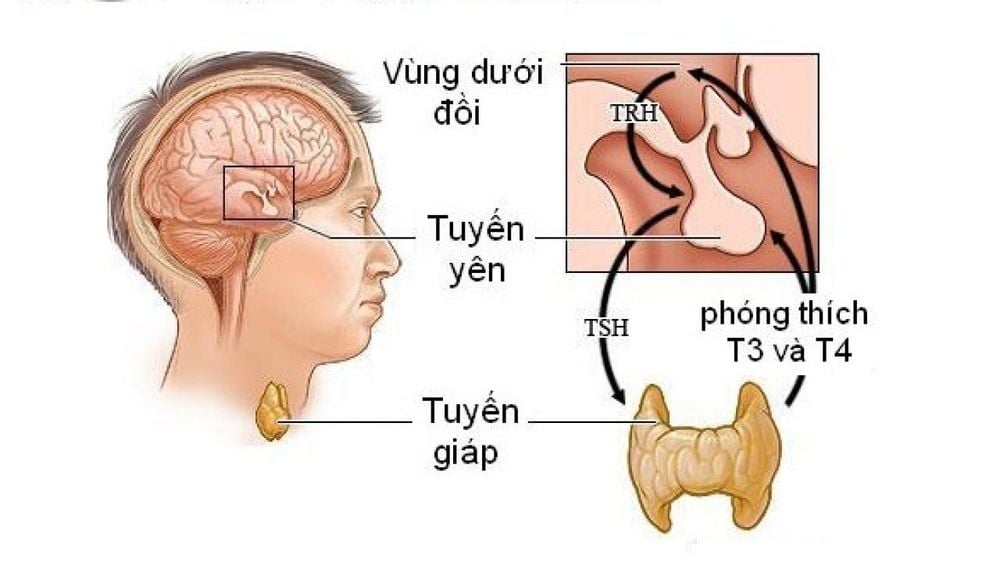
Much of the role the amygdala performs is accomplished through its primary function: regulation of the hypothalamus through various neural pathways.
1.2. The nucleus of the amygdala The amygdaloid body, or just the amygdala, is the subcortical gray matter of the limbic system supplied with blood by the anterior choroidal artery. It contains 13 nuclei grouped into three functionally different nuclear divisions:
Basolateral groups Central groups Corticoid medial groups 1.3. Basal (abdominal) group This group includes the lateral nucleus, basal nucleus, and minor basal nucleus. It is larger and phylogenetically newer. Functionally, it belongs to the limbic system. It has reciprocal connections with sensory-associated areas of the cerebral cortex, all four of them (auditory, visual, and auditory cortex).
It projects to three different places:
Median dorsal nucleus of the thalamus Basal nucleus (by Meynart) ventral striatum This nucleus regulates various functions of the thalamus, such as:
Coping with stress Somatic and autonomic reflexes Feeding and drinking behavior 1.4. Biomedical group (dorsomedial) This group contains the cortical nucleus and the nucleus of the lateral olfactory tract. Compared with the basal group, it is smaller, phylogenetically older, and functionally part of the olfactory system. It is actually where the olfactory fibers end from the olfactory bulb. This nucleus projects to the ventromedial nucleus of the hypothalamus and it plays a role in behaviors related to hunger and eating.
1.5. Centromedial Group This group is composed of intermediate and central nuclei. It is actually a place where both the lateral and external nuclei of the body project. This nucleus is interconnected with visceral sensory nuclei and autonomic nuclei in the brain stem that are involved in respiratory and cardiovascular functions.
In addition to these nuclear groups, there is a separate nuclear group that cannot be easily classified into any of these three groups and is therefore listed separately. These include interstitial cell masses and the hippocampus amygdala.
1.6. Pathways Each amygdaloid nucleus receives input and sends output to many distinct regions of the brain. These inputs and outputs are called pathways, and depending on whether they are received or sent, these paths can be radial and directed.

Amygdala là một trong hai nhóm nhân hình quả hạnh nằm sâu trong thùy thái dương
In any case, it is clear that all these fibers transmit a broad spectrum of auditory, visual and optical information to the amygdala. For the reason that many afferent fibers are received, the basal nucleus group is often referred to as the amygdaloid region of sensory convergence.
All these pulses are received to the basal group which is then projected to the focal group where they are supposed to be aligned and then sent to different motor centers to stimulate a response to the received information Okay. For that reason, the central group is often referred to as the amygdaloid region of motor divergence.
Specifically these are the structures that send fibers to the amygdala:
Smell Prefrontal cortex Cingulate gyrus Basal forebrain Intermediate thalamus Hypothalamus Brain stem Effective prediction It was previously mentioned that The main role of the amygdala is to regulate hypothalamic activity. It does so through two main pathways: the striatal terminal pathway and the ventral amygdalo fungal pathway.
The stria terminalis This pathway runs from the central nucleus of the amygdala to the ventricular nucleus of the hypothalamus. This pathway also carries fibers to the septal nuclei and to the hippocampal regions of the brain.
Along with these accessory fibers, the terminal striatum carries projection fibers from these three areas back to the amygdala. Stria terminalis plays an important role in various types of organism responses to stress.
When it leaves the central nucleus, most of its fibers lie between the caudate nucleus and the thalamus, followed by the vena cava. For its entire length, the striatum is related to a group of neurons called the bed nucleus of the striatum, the function of which will be described later.
The terminal fibers of the terminal striatum of the striatum are spreading as multiple rays and they reach various nuclei of the hypothalamus, such as the ventral nucleus, the anterior nucleus, the anterior nucleus, and the lateral hypothalamic nucleus. They also form synapses with neurons of the nucleus accumbens, septum nucleus, caudate nucleus, and stuffed nucleus. Since it is connected to the bone marrow via the septal nuclei, the striatum terminals indirectly regulate the brain stem.
The aforementioned striatal striatal nucleus is a forebrain structure responsible for receiving projections (among other areas), from the basal lateral amygdala and in turn projecting to target areas hypothalamus and brain stem, mediate many autonomic and behavioral responses to hostile or threatening stimuli. It is a morphologically different structure in the male brain than in the female brain, which is twice as large in males as in females. For that type of nucleus, we say that they are sexually dimorphic.
Different morphology of the bed nucleus of the striatum between males and females also determines specific differences when it comes to phobias. This is why women are said to be more fearful than men. Other microfinance terminal functions correlate with threat response anxiety. The bed nucleus of the striatum is thought to act as a relay site in the hypothalamic-pituitary-adrenal axis and modulate its activity in response to acute stress lasting more than 10 min. It is also thought to promote behavioral inhibition when encountering unfamiliar individuals.
Amygdalofugal pathway This pathway is the main pathway of the amygdala. It originates from the central nucleus and tripartite nucleus of the amygdaloid complex and carries fibers to many locations in the nervous system.
Axons from the basal group are mediated by the renewer, and terminate in the hypothalamus and septal nucleus. It is well known that the regenerative agent projects cholinergic neurons to the cerebral cortex important for the regulation of social behavior. In addition, the basal group projected diffuse fibers and synapses with broad regions of the prefrontal and frontal cortex, gyrus, medial and temporal cortex. On the other hand, fibers from the central nucleus are directed longitudinally toward the brain stem, to terminate in the raphe nucleus, along with other structures such as the periorbital gray matter and the orbital coeruleus. These structures also send fibers. their return to the amygdala.
The amygdalo fungal and stria terminalis pathways combine to allow the amygdala to directly control the medial hypothalamus, lateral hypothalamus, and periventricular gray. This pathway is especially important for associative learning.

Vai trò chính của hạch hạnh nhân là điều chỉnh hoạt động của vùng dưới đồi
2. Damage to the amygdala
Knowing the functions of the amygdala is very important, because if your patient has some type of lesion in the temporal lobe, you will easily notice if the amygdala is also affected. People with damaged amygdala show symptoms of so-called Klüver-Bucy syndrome. This syndrome is defined by the following symptoms:Inability to visually perceive surrounding objects Tends to check surrounding objects by smelling or chewing them Can't resist the need to explore surroundings and overreacts to visual stimuli Expresses excessive fear and anger Eating unusual amounts of food even when the patient is not hungry Along with these symptoms, amygdala damage can may be followed by amnesia, amnesia, and aphasia.
If you have a need for consultation and examination at Vinmec Hospitals under the national health system, please book an appointment on the website (vinmec.com) for service.
Please dial HOTLINE for more information or register for an appointment HERE. Download MyVinmec app to make appointments faster and to manage your bookings easily.
Reference sources: kenhub.com, healthline.com


