This is an automatically translated article.
Posted by Doctor Mai Vien Phuong, Department of Medical Examination & Internal Medicine - Vinmec Central Park International General Hospital
Double esophageal cyst is the second most common type of gastrointestinal duplication cyst, after ileal double cyst. However, less than 100 cases of double esophageal cyst are currently documented. Double esophageal cyst usually occurs alone; however, two cases of multiple (two) double esophageal cysts have been reported.
1. Symptoms of Double Esophageal Cysts
Most esophageal cysts do not have debilitating symptoms. Affected children may complain of nonspecific symptoms involving the esophagus or the respiratory tract, depending on size and presence, or in the absence of hemorrhagic, infectious, ruptured, or related complications. to the respiratory tract. Double esophageal cysts may be asymptomatic and incidentally discovered during thoracotomy for other surgical conditions. Some patients present with symptoms associated with obstructive pneumonitis due to the large compression of the tracheal tree by a double esophageal cyst, which is not different from the symptoms of common pediatric mediastinoma.
The radiographic findings of double esophageal cysts are generally nonspecific and similar to those of other common noncancerous or mediastinal cancers. Contrast and non-contrast computed tomography can provide information about esophageal cysts and adjacent esophagus and tracheal tree. If computed tomography is equivalent, MR imaging and endoscopic transesophageal ultrasound (endoscopic ultrasound) can be used for preoperative evaluation and preoperative diagnosis of double esophageal cysts. . T1-weighted magnetic resonance imaging can accurately visualize an esophageal cyst, even in the presence of bleeding and infection complications [19]. Endoscopic ultrasonography is considered an effective adjunct to the X-ray and MR evaluation of esophageal cysts, and often reveals oesophageal double cysts as anechoic or hypoechoic cysts. Endoscopic ultrasound-guided fine-needle aspiration biopsy can provide a pathological diagnosis, helping to differentiate double esophageal cysts from other mediastinal tumours. Double esophageal cyst may occur concomitantly with esophageal stricture/atony, tracheal fistula, and other tracheal/pulmonary malformations. Preoperative esophagography and endoscopic ultrasonography can help rule out coexisting malformations and outline the surgical plan.
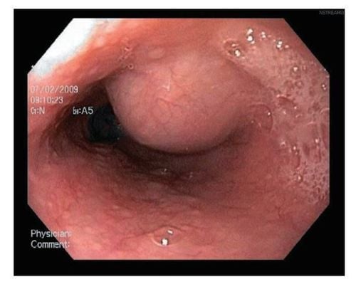
Hình ảnh nang thực quản đôi được nhìn thấy trên nội soi
2. Diagnostic criteria for double esophageal cyst
Histology provides the best diagnostic tool of double esophageal cyst. The diagnostic criteria for a double esophageal cyst are as follows: the tumor is attached to the esophageal wall, communicates with the epithelium of gastrointestinal origin, and is lined by two layers of stromal muscle. Double esophageal cysts may be lined by pseudomembranous columnar epithelium, gastric mucosal epithelium, and squamous epithelium. The cyst wall may also contain cartilage tissue. Ectopic gastrointestinal mucosa may occur independently or simultaneously with esophageal cysts in the upper and/or middle portions of the esophagus. The presence of gastric mucosa in the lower part of the esophagus is commonly referred to as Barrett's esophagus. These ectopic gastric mucosa typically present as small patches of columnar epithelium containing gastric glands, which vary in size from millimeters to centimeters on endoscopy and are detectable by pertechnetate technetium scanning. -99 meters. Only one case of double esophageal cyst with ectopic gastric mucosa in an infant presenting with hemoptysis has been reported. The respiratory bleeding in this patient may be due to the erosive effect of gastric acid secreted by the ectopic gastric mucosa. Ectopic pancreatic tissue has also been reported to co-occur with esophageal cysts. Similar to gastric mucosa, ectopic gastric mucosa is still at risk of malignancy.
3. Treatment of esophageal cysts
Surgical treatment to remove the entire double esophageal cyst is indicated if the cyst has complications such as inflammation, bleeding ulcer, perforation, even gastrointestinal cancer. Surgical resection is the mainstay of treatment for symptomatic double esophageal cysts. This procedure can be done through a thoracotomy or thoracoscopy to remove a double esophageal cyst. Complete removal of the double esophageal cyst is essential to minimize the risk of recurrence. Endoscopic thoracoscopic resection is believed to be as effective as the conventional open procedure, and offers additional minimally invasiveness and rapid postoperative recovery. Complete or partial endoscopic resection after endoscopic ultrasound evaluation Endoscopic ultrasound has also been reported to be an effective and safe treatment for small-sized double esophageal cysts. Small size, vascular and surface layer. Small, asymptomatic double esophageal cysts can be monitored without intervention. A previous study showed that esophageal cysts were associated with mild upper gastrointestinal symptoms, and in such cases, the size of esophageal cysts remained unchanged on Endoscopic Ultrasound for more than 13 years. If necessary, small double esophageal cysts can be removed using endoscopy or percutaneous anhydrous ethanol injection.

Cắt bỏ bằng phẫu thuật là phương pháp điều trị chính cho nang thực quản đôi có triệu chứng
Conclusion
Double esophageal cyst is an uncommon differential diagnosis in children with posterior mediastinal cystic mass. Vinmec International General Hospital uses a combination of medical imaging techniques, such as contrast oesophagography, computed tomography scan, magnetic resonance imaging, and endoscopic ultrasound, which can help identify specific characteristics. The score of the double esophageal cyst relates to its adjacent anatomy and excludes possible concomitant tracheal malformations. Ectopic gastric mucosa can be detected retrospectively in double esophageal cystectomy specimens. Large and/or symptomatic esophageal cysts should be treated surgically, while small and/or asymptomatic esophageal cysts should be monitored closely, especially if the cyst is located near the upper chest or where the trachea bisects.
To learn more about esophageal cysts, you can refer to the article: Overview of esophageal cysts
Please dial HOTLINE for more information or register for an appointment HERE. Download MyVinmec app to make appointments faster and to manage your bookings easily.
References:Zhefeng Zhang et al., Double esophageal duplication cysts, with ectopic gastric mucosa: a case report. Journal of Cardiothoracic Surgery volume 8, Article number: 221 (2013) Emura T, Hashizume K, Asashima M. Experimental study of the embryogenesis of gastrointestinal duplication and enteric cyst. Pediatr Surg Int. 2003;19:147–151. [PubMed] [Google Scholar] Iyer CP, Mahour GH. Duplications of the alimentary tract in Children and children. J Pediatr Surg. 1995;30:1267–1270. doi: 10.1016/0022-3468(95)90482-4. [PubMed] [CrossRef] [Google Scholar] Kiratli PO, Aksoy T, Bozkurt MF, Orhan D. Detection of ectopic gastric mucosa using 99mTc pertechnetate: a review of the literature. Ann Nucl Med. 2009;23:97–105. doi: 10.1007/s12149-008-0204-6. [PubMed] [CrossRef] [Google Scholar]