This is an automatically translated article.
Tumors of the pharynx account for less than 1% of all head and neck tumors. These tumors can be benign or malignant, they can originate from nerves, salivary glands or are metastatic lesions,... Surgery to remove the tumor on the side of the pharynx is the main treatment. for these cases.
1. What is a cavity tumor in the throat?
Pharyngeal cavity tumors are tumors arising and growing within the anatomical structures of the lateral pharynx. The nature of the tumor can be benign such as neurofibromatosis, salivary gland tumor, carotid tumor, cystic tumor.
Tumor can also be malignant such as salivary gland cancer, metastatic lymphoma, carcinoma, sarcoma. Tumors in the side of the pharynx are not common, but they are important because of their anatomical location around the cervical nerve vascular axis and the base of the skull.
Parapharyngeal tumor resection is an operation to widen the access and surgical field of the lateral pharynx to dissect, resect the tumor and restore its structure.
There are many routes into the lateral pharynx such as: oropharyngeal line, anterior and posterior border of the sternocleidomastoid muscle. However, the safest, most widely used and most accessible way to completely resect the tumor in the lateral pharynx is the lateral cervical line (Sébileau-Carrega).
2. Indications and contraindications of lateral pharyngeal tumor resection
Nasopharyngeal resection is indicated in cases where the tumor in the side of the pharynx can be benign or malignant:
Tumor of the parotid deep lobe Fibroids Neurofibromas Cysts Lymphomas Tumors Blood Tumor tumor metastasis to lateral pharyngeal lymph node and Cuneo-Krause lymph node (posterior septal foramen). Fetal muscle sarcoma. Tumor resection in the pharynx is contraindicated in the following cases:
Tumors have destroyed the base of the skull or have invaded the vascular axis or posterior foramen. Patients with systemic diseases such as diabetes, cardiovascular disease, blood pressure (not an absolute contraindication).

Phẫu thuật cắt u trong họng chống chỉ định với các bệnh nhân có các bệnh lý toàn thân như đái tháo đường, tim mạch, huyết áp
3. Steps to perform surgery to cut the tumor on the side of the throat
3.1. Step 1: Check patient records The doctor checked the medical record before anesthesia and checked the blood type3.2. Step 2: Check the patient
Check the patient's condition, including checking: Pulse, temperature, blood pressure.3.3. Step 3: Carry out surgery
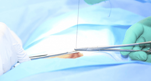
Stage 1: The surgeon makes a lateral neck incision
If the tumor is located in the lower part (medial and lower extremities) of the lateral pharynx, the incision will extend from the level of the posterior plate to the angle of the jaw. If the tumor is located high (upper pole) and deep in the lateral space of the pharynx, an incision is made according to the Sébileau-Carrega line. Stage 2: The doctor dissects the superficial cervical fascia and the middle cervical fascia along the skin incision to penetrate the carotid trough. During this procedure, the doctor must tie and ligate the branches of the external jugular vein. Stage 3: The doctor finds and ligation of the facial vein, it is necessary to find the external jugular vein, the 12th cranial nerve, and the facial vein (Farabeuf's triangle), to be able to widen the access to the submandibular gland. Stage 4: The doctor removes the submandibular gland to widen the access to the lower pole of the lateral pharynx. Then, the doctor will dissect the submandibular capsule, ligation, and remove both the duct and the gland.
5: The doctor performed a dissection through the middle and deep cervical fascia to expose the tumor capsule of the lateral pharynx, well stopping the blood vessels feeding the capsule with bipolar electrocoagulation. The superior tumor pole can often adhere to the fascia, basilar periosteum, or carotid bay, which should be taken into account.
Tumor dissection can be done by hand, trowel or surgical forceps. Tumors such as cysts, solid tumors, neurofibromas, salivary gland tumors, and lymph nodes are often less adherent. But carotid tumor resection will have to be very careful both to separate the tumor and to coagulate the tumor to remove the tumor from the vascular and nerve sheath. Once the tumor has been separated, it is easy to remove, but the incision can be quite large.
6: The doctor checks the surgical cavity, stops bleeding carefully, puts gelaspon if necessary, places a closed drainage. Then close the surgical cavity with 2 or 3 layers including deep cervical fascia, muscle, subcutaneous tissue and skin; Apply gentle pressure to the posterior corner of the jaw and neck.
After surgery to remove the tumor on the side of the pharynx, the patient should be closely monitored. The patient can have sutures removed after 7 days, when it is safe for surgical scarring.

Bệnh nhân có thể được cắt chỉ sau 7 ngày, khi đã an toàn về sẹo hóa hốc mổ.
4. Possible complications in lateral pharyngectomy and treatment
4.1. Anesthesia accident
During surgery, it is necessary to pay attention to complications of breathing tube collapse, pneumothorax.
4.2. Bleeding accident
During the surgery to remove the tumor in the throat, there may be bleeding due to the small artery surrounding the tumor capsule. More severe bleeding events often occur in cases where the tumor has spread to the base of the skull, with tumor feeding branches arising from the internal carotid artery.
The tumor must be completely removed to stop bleeding. Careful electrocoagulation should be performed on the tumor attachment area and the tumor feeding branches. At the same time, it is necessary to consider and evaluate the amount of blood the patient has lost for blood transfusion, to compensate for blood accordingly. Must closely monitor the parameters of pulse, blood pressure, of the first class nursing regimen for bleeding cases.
4.3. Paralysis of the nerves of the pharyngeal plexus, the X nerve and the cerebrospinal fluid leak
Cerebrospinal fluid leak occurs when the tumor has spread to the base of the skull and has destroyed the bone at the base of the skull. In these cases, surgery is required to seal the craniofacial cleft.
Throat resection surgery is a complex surgery and requires high expertise and skill from the performer. Currently, surgery is being performed routinely at Vinmec International General Hospital. In addition, at Vinmec, we also receive examination and treatment of common nasopharyngeal diseases, tumors of the head, face and neck, congenital malformations of the ear, nose and throat area with the most optimal internal - surgical methods for patients. patients, both children and adults.
Coming to Vinmec International General Hospital, patients will receive a diagnosis with the leading modern medical equipment in the country, combined with direct and dedicated examination from a team of qualified medical staff. Professional, experienced, help the treatment and recovery process become faster.
Please dial HOTLINE for more information or register for an appointment HERE. Download MyVinmec app to make appointments faster and to manage your bookings easily.


