This is an automatically translated article.
Tumor of the orbital top is one of the very dangerous eye diseases, causing damage to vision, if not detected and treated in time, can lead to diverse and complicated complications.
In addition, due to the specific anatomical location, orbital tumors are often detected late, only when the tumor has spread widely and seriously affects the patient's vision and health. Often the treatment of choice for many patients is surgery for apical orbital tumors.
1. What is surgery for apical orbital tumor?
Symptoms of orbital tumor
The most common symptoms of orbital tumor are protrusion, pain in the orbit, crossed eyes, drooping eyelids and visual impairment.
Except in exceptional cases, usually tumors at the top of the orbit tend to take up space and push the eyeball posteriorly. For example, optic nerve tumors tend to push the eyeball in an anterior longitudinal direction, while lacrimal gland tumors tend to push the eyeball downward and inward. In general, the most obvious and prominent sign of an orbital tumor is the protrusion of the eye. Observing the convex orientation of the eyeball can help make relative inferences about the location of the tumor. In addition to clinical examination, CT scan is also very important in diagnosing the size, nature, location, invasion... of the orbital apical tumor.
Surgery for apical orbital tumor is considered the most effective treatment modality in the majority of cases. The choice of incision depends on the location and size of the tumor, but for tumors at the top of the orbit, the opening of the skull cap (forehead or keyhole area) is often chosen because it is for surgery. wide, allowing doctors easy access to the top of the orbit and to the base of the skull (especially for skull base tumors that invade the orbit or vice versa).
Indicated cases
Surgery to remove the tumor (for tumors with symptoms such as eye protrusion or progressive visual impairment). Surgical decompression of the optic nerve (with large invasive tumors not capable of radical surgery). Surgical biopsy to remove tumor samples for diagnosis. Cases of contraindications
Severe systemic condition: Metastatic cancer in the orbit... Eyes no longer functional (dry cornea, retinal detachment...).

U đỉnh hốc mắt gây lác mắt
2. Preparation for surgery
Before surgery, the patient will have a careful eye exam, CT scan to determine the location, size and other properties of the mass, evaluate the protrusion, drooping eyelids, congestion, visual acuity. , fundus condition, eye muscles...
In addition, the patient may also be asked to perform endocrine tests to help diagnose Basedow's protrusions, blood count tests to help differentiate the condition. pseudotumor inflammation ... or cerebral angiography to exclude lesions such as carotid artery - cavernous sinus .
The composition of the operating team includes 7-8 people including anesthesiologists, neurosurgeons, surgical assistant doctors, anesthesiologists, nurses serving surgical instruments, nurses run outside.
Surgical instruments include head mount (Mayfield frame), microsurgery instrument, Sonopet ultrasonic scalpel, hemostatic device, machine drill (cutting + grinding), nerve positioning system (Neuro Navigation), microsurgical glasses, consumables such as cotton, gauze, skull wax, Surgicel, Prolene 4.0 thread, Vicryl 0...
3. Surgical procedure for apical orbital tumor
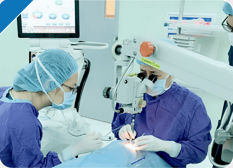
When starting surgery for apical orbital tumor, the patient is placed in the supine position, with the head fixed on the head (Mayfield frame), then endotracheal anesthesia is carried out.
Steps of surgery for apical orbital tumor
Step 1: Register the nerve positioning system (Neuronavigation) Step 2: Widely disinfect the surgical area, anesthetize the incision area (on the brow or forehead hairline) ). Step 3: Make skin incision, separate muscle fascia, periosteum to reveal skull bone. Step 4: Open the skull cap by drilling machine, open dura, subarachnoid space to aspirate less cerebrospinal fluid to flatten the brain. Step 5: Place the valve of the brain (if necessary), put on the microsurgery glasses. Step 6: Identify nerve number II, carotid artery area Step 7: Use a drill to grind bone at the top of the eye socket, expand it with the trigger. Step 8: Surgically separate the tumor from the nerves and blood vessels (if necessary). Partial removal of the tumor with microsurgery scissors or ultrasound knife. Send a portion of the tumor sample for immediate biopsy to confirm the diagnosis and give appropriate treatment. Step 9: During surgery, use Navigation to determine the position of the correlation between the tumor and the nerves and blood vessels. Stop bleeding. Step 10: Close the dura, suspend the dura, put the bone back. Place surgical drainage if necessary, close the incision.

Phậu thuật u đỉnh hốc mắt cần được thực hiện theo đúng quy trình
4. Post-operative monitoring and treatment
Postoperative follow-up:
Observe the whole body condition: Perception, pupil size, round or distorted shape, reflectivity to light, breathing capacity, temperature.... Bleeding at the site surgery. Drainage (if applicable): Drainage is usually withdrawn after 48 hours. Common complications and treatment methods
Postoperative bleeding: the surgical process may inadvertently affect large arteries or small blood vessels... leading to postoperative bleeding. In this case, it is possible to manage according to the bleeding lesion and consider surgical removal of the hematoma if necessary. Infection: conduct medical treatment (change dressing, keep absolute sterility, prescribe antibiotics). Soft swelling around the eyes: Apply warm compresses to help relieve swelling, medical treatment (anti-inflammatory)... Decreased vision: Continue to monitor, medical treatment (if necessary). Customers who need medical examination and treatment can directly go to Vinmec Health System nationwide or contact to book an appointment online HERE.
MORE:
Brain abscess: When to operate? How is traumatic brain injury surgery? Treatment in patients with cerebral hematoma due to traumatic brain injury
Notes on radiation therapy for cancer


