This is an automatically translated article.
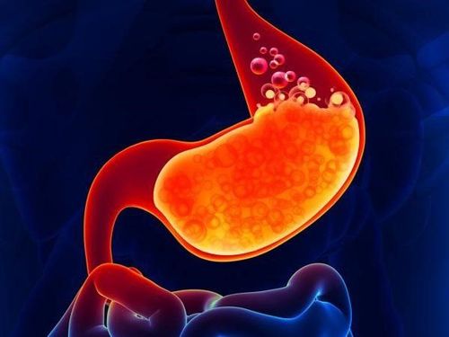
Stomach is one of the most important internal organs of the body. Stomach anatomy helps readers better understand the position, the parts that make up the stomach.
1. Stomach position
The stomach is the largest part of the alimentary canal, the upper part connects to the esophagus through the foramen, and the lower part connects to the duodenum through the pyloric orifice. The stomach is an intraperitoneal organ located in the upper layer of the transverse mesentery, the epigastrium, and the left subdiaphragmatic cell.The basic function of the stomach is to store and digest food. This is also known as the first part of the small intestine. When compared to the abdominal wall, the stomach belongs to the epigastrium, the left hypochondrium and the navel. The stomach is J-shaped, but with good elasticity, this organ has a shape that changes depending on the position, the time of examination, whether it is containing food or not,... The volume of the stomach is about 2 - 2.5 liters or more.
2. Stomach Anatomy
Physiological anatomy of the stomach consists of the following parts:
2.1 Gastric anatomy: External appearance The stomach has 2 sides, the front and the back. The stomach has 2 borders: the left large gastric curvature, with a cardiac defect separating the fundus from the esophagus; The right minor curvature has an angular defect that is the boundary between the body and the subject matter.
In terms of external appearance, the stomach is divided into the following parts:
Cardiac: The area is about 5 - 6cm2, with a cardiac hole connecting to the esophagus. The orifice of the stomach has no sphincter or valve, only a mucosal fold separating the stomach from the esophagus; fundus: Located above the plane through the cardial foramen, it normally contains air; Body: The part of the stomach below the fundus, the lower limit is the oblique plane passing through the angle defect. The body contains glands that secrete Pepsinogen and Hydrochloric Acid (HCl); Pylorus: Consists of a funnel-shaped pyloric cave that secretes Gastrine and a part of the pyloric canal with well-developed muscles. The pylorus is located to the right of the 1st lumbar vertebra, with the pyloric foramen connected to the duodenum. The pyloric orifice has 1 sphincter. When this muscle is enlarged, it can cause hypertrophic pyloric spasm, which is common in neonates.
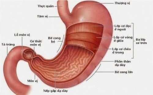
Hình ảnh giải phẫu dạ dày
2.2 Anatomy of the stomach: Internal shape The internal structure of the stomach consists of 5 layers: serosa, subserosal plate, muscle layer (3 smaller muscle layers are: longitudinal muscle, diagonal muscle, sphincter), layer submucosa and mucosal layers. The characteristics of the layers in the stomach are as follows:
Serous layer: Located in the outermost layer, belonging to the visceral peritoneum; Subserosal plate: A very thin connective tissue, almost adhered to the muscle layer, except for the part near the 2 sides of the gastric curvature; Muscular layer: The layer adapted to the mixing of food in the stomach. The muscular layer is composed of: longitudinal muscle (continuous with longitudinal muscle fibers of the esophagus and duodenum, thickest, running along the small gastric curvature), sphincter (encloses the entire stomach, especially in the stomach). pylorus), oblique muscle (incomplete layer, running around the fundus, diagonally downward toward the greater curvature of the stomach); The submucosa (submucosal sheet): It is a very loose connective tissue, easily pushed; Mucosal layer: The inner lining of the stomach. The stomach is fed by an artery originating from the visceral body, forming two arcs: a large arc running along the great curvature, and a small arc running along the small curvature. The characteristics of the gastric artery are as follows:
Small curvature of the gastric artery: It is formed by the right gastric artery (branch of the hepatic artery) and the left gastric artery (branch of the visceral artery). In addition, there are short gastric arteries, arteries for the cardia and esophagus, and posterior basal artery; The great curvature of the gastric artery is formed by the right gastric omental artery (branch of the gastroduodenal artery of the common hepatic artery) and the left gastroocardium (belonging to the splenic artery). In addition, there are short gastric arteries, body arteries,... Nervous system: The stomach is innervated by the vagus nerve and some branches of the medulla (parasympathetic part). The sympathetic part consists of sympathetic fibers from the lumbar and thoracic sympathetic ganglia.
Gastric lymph nodes: Drained into 3 groups: Gastric lymph nodes (located along the small curvature), gastric lymph nodes - omentum (located along the great gastric curvature) and other gastric lymph nodes. Pancreatic splenic lymph node (located in the splenic omentum).

Dạ dày là một trong những cơ quan nội tạng quan trọng nhất của cơ thể
3. Stomach Function
According to gastric anatomy experts, this organ performs 4 main functions including:
Digestive function: HCl acid in the stomach has the effect of activating digestive enzymes, regulating the opening/closing of the pylorus, stimulates pancreatic secretion. Mucus plays an important role in protecting the mucosa from damage by gastric juice. Besides, Pepsinogen and HCl help to break down proteins into polypeptides; Endogenous factors help absorb vitamin B12. The stomach also produces secretin - a hormone that stimulates the secretion of pancreatic juice; Motor function: Gastric tone, intragastric pressure about 8-10cmH2O. This pressure is generated by frequent contractions of the muscular layer in the stomach; Excretion function: On average, the stomach excretes about 1-1.5 liters of gastric juice per day, plasma proteins (albumin, immunoglobulins), enzymes pepsinogen, pepsin, glycoproteins, endogenous factors and acids; Peristalsis function: It takes about 5 - 10 minutes for food to enter the stomach before the stomach begins to have a peristalsis. Peristalsis begins in the middle part of the stomach body, the closer to the cardia, the stronger and deeper the peristalsis. Then, every 10 - 15 seconds, there will be a repeating peristaltic wave. After that, the process of mixing food with gastric juice takes place, and at the same time, the food is crushed and ejected into the intestines. The article helps readers have an overview of stomach anatomy and important functions of this organ for the body. Therefore, each person should maintain a healthy diet, rest and reasonable physical activity to keep the digestive system and stomach healthy. If there are signs of suspicion of stomach disease, the patient should go to the doctor, diagnose and treat early.
Please dial HOTLINE for more information or register for an appointment HERE. Download MyVinmec app to make appointments faster and to manage your bookings easily.


