This is an automatically translated article.
The article is professionally consulted by Master, Doctor Le Xuan Thiep - Radiologist - Department of Diagnostic Imaging - Vinmec Ha Long International Hospital. The doctor has extensive experience in the field of diagnostic imaging.RT-PCR is currently the leading tool to diagnose COVID-19 and is also a technique to make a definitive diagnosis. However, many studies around the world also show that computed tomography is indispensable in supporting the diagnosis, care and monitoring of Covid-19 patients.
1. RT-PCR technique to diagnose Covid-19
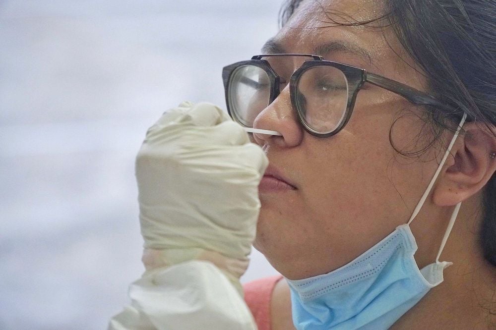
Covid-19 caused by the SARS-CoV-2 virus is a global pandemic with millions of people infected and hundreds of thousands of deaths worldwide. Testing is now an important tool to help diagnose and control the spread of disease in the community.As recommended by the World Health Organization WHO, RT-PCR (Real Time Polymerase Chain Reaction) is the most optimal method to diagnose Covid-19. This method helps to detect the nucleotide acid of the SARS-CoV-2 virus in patient samples taken from the nasal cavity, oropharyngeal cavity or bronchoalveolar lavage of suspected infected people.
The working principle of RT-PCR is to make a large number of copies of DNA from a selected segment of DNA in a short time. DNA replication takes place in an in vitro environment similar to that during cell division. By RT-PCR technique, from a small amount of DNA in the patient sample will be accurately amplified into a large amount up to millions of copies to serve the investigation process in the reaction.
RT-PCR technique for COVID-19 diagnosis gives more accurate and faster results than traditional testing methods. However, in practice, there are many cases of RT-PCR test giving false negative results, especially when the patient has an early infection. The cause may be due to substandard swab swab or test sensitivity is not guaranteed. Therefore, the combination of imaging diagnostic tools such as computed tomography to support the diagnosis, care and monitoring of the progress of Covid-19 patients is extremely necessary.

Kỹ thuật RT-PCR chẩn đoán COVID-19 cho kết quả chính xác và nhanh hơn các phương pháp xét nghiệm truyền thống
2. The role of computed tomography in the diagnosis of COVID-19
Computed tomography is a technique that uses X-rays to scan an area of the body, then coordinates with a computer to produce two or three-dimensional images of the location to be taken. The advantage of computed tomography is that the images are clear and there is no overlap. Clear image resolution makes diagnosis many times more accurate than X-ray methods.In the diagnosis of Covid-19, with the ability to perform cross-sections of less than 1mm thickness and clear polyhedral rendering, computed tomography helps to detect specific lung lesions in pneumonia caused by Covid-19. Reports have shown that computed tomography has a sensitivity of 56-98% for detecting Covid-19 pneumonia even in the early days of disease onset (while in the early stages, the sensitivity of RT- PCR is only from 60-71%.
Another study conducted in Japan found that computed tomography helped detect lung damage in 54% of cases of Covid-19 infection but did not have any signs of onset. Thanks to that, the outstanding advantages that computed tomography help to correct early diagnosis for patients with false negative results in RT-PCR test, greatly benefit the treatment and prevention of epidemics.
On computed tomography images, the lungs of Covid-19 patients have the following prominent features:
Multifocal lesions distributed in the periphery, subpleural and bottom lung. Lesions occur on both sides of the lung. Lesions presented by opacoid opacities range from simple opacoid opacities in the early stages, to opacular lesions with interlobular septal thickening, medial lobular wall thickening, reticular thickening, and opacular opacular opacities. cloudy glass, with partial thickening of the lung; simple solid pulmonary opacities. Another type of lesion is the band opacity, but this is less common. The blurs are usually geometric, in some cases circular or inverted halos. Some other characteristic signs are: the vascular structure inside the opacities is enlarged, the interlobular septum and in the lobules are thickened, creating irregular paved images, bronchioles containing air,... Types of lesions such as nodular lung lesions, pleural effusion, hilar lymph nodes, mediastinal nodes are not specific for Covid-19. Therefore, if these lesions are present, pneumonia of another etiology should be considered.
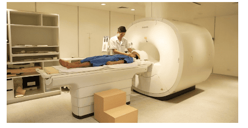
Ưu điểm của chụp cắt lớp vi tính là cho hình ảnh rõ nét và không có hiện tượng chồng lên nhau
3. Diagnosis of Covid-19 progression by computed tomography
Studies have shown that in mild Covid-19 patients, without respiratory failure, without mechanical ventilation, the course of the disease corresponds to the change on computed tomography:Early stage (0) -4 days): On computed tomography scans, it is mainly a group of blurred glass lesions, with a small lesion area. Advanced stage (5-8 days): The patient's inflammatory state is widespread and more severe, the expression on computed tomography is from the expression of the opacified glass lesions appearing with the presence of paved stones. uniform and dense lungs. Peak phase (9-13 days): The rate of spread of the lesion slows down, on computed tomography, solid pulmonary imaging predominates alongside diffuse opacities and paved imaging patterns. irregular, appearing fibrous bands in the parenchyma. Absorption phase (more than 14 days): Inflammation is controlled during this period, solid lung lesions are gradually absorbed to medial opacities with no irregular paved pattern, then then this sign is erased or fibrous bands appear. The evolution of severe Covid-19 cases on computed tomography images is:
Lesions increase in number, distribution is more and more diffuse, the number of affected lung lobes is more. The lesion increased in size, at this time the lesion was not only localized in the subpleural periphery but also gradually spread towards the center. In terms of density, the lesion gradually increases, with the opaque glass type, the density gradually increases in the direction of forming an irregular stone-paved cloud, dense lung tissue gradually replaces the opaque area. With solid lung lesions, the density is also increased, creating a white lung image. Lung function is severely impaired. The disease often worsens in elderly patients with many underlying medical conditions.
Các nghiên cứu đã chỉ ra ở những bệnh nhân Covid-19 thể nhẹ, không có suy hô hấp
4. Differential diagnosis of Covid-19 from other diseases by computed tomography
Studies have shown that computed tomography images can distinguish pneumonia lesions caused by Covid-19 from pneumonia caused by other viruses as follows:Pneumonia due to Covid-19 has a distribution of lesions in High peripheral opacity, superior opacity, fine reticular opacity is more common, vascular wall thickening is more, reversed halo sign is more common. Pneumonia due to Covid-19 rarely has pleural effusion and rarely lymphadenopathy. Most studies show no cavernous lesions in patients with Covid-19. Compared with lung injury caused by Corona virus SARS (acute respiratory syndrome in 2002) and MERS (acute respiratory syndrome in the Middle East in 2012), in patients with Covid-19, Halo sign and reversed Halo sign were found. more than. Lung injury caused by SARS and MERS is predominantly a single focal point. Thus, the RT-PCR technique to diagnose COVID-19 is the current gold standard, but the use of computed tomography is indispensable in supporting the diagnosis, care, and monitoring of the disease. Covid-19 patients.
Please dial HOTLINE for more information or register for an appointment HERE. Download MyVinmec app to make appointments faster and to manage your bookings easily.
MOREWhat is the source of 2019-nCoV infection? How does the 2019-nCoV virus spread? Is rubbing eyes and nose contagious? Is 2019-nCoV the same as the virus that causes MERS and SARS?


