This is an automatically translated article.
The article was written by Dr. Trinh Van Dong - Radiologist - Department of Radiology - Vinmec Ha Long International General Hospital.Currently, there are many methods used to diagnose pulmonary embolism. In particular, pulmonary embolism electrocardiogram is being widely used in the detection and treatment of pulmonary embolism.
1. What is an electrocardiogram?
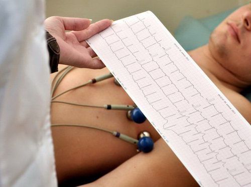
Electrocardiogram (English name is Electrocardiogram - ECG for short) is a type of graph that records the electrical signals of the heart. This test is widely used to detect problems and monitor the condition of the heart, especially to detect heart diseases such as arrhythmia, myocardial infarction, heart failure, etc. ..Electrocardiogram is a non-invasive, painless test with fast results. In an electrocardiogram, electrodes are attached to the chest (sometimes to the legs) to capture the electrical signals of the heart and are recorded by an electrical recorder.

Các điện cực được gắn vào ngực để đo và ghi lại bằng máy điện
2. Electrocardiogram in the diagnosis of pulmonary embolism
2.1 What is a pulmonary embolism? A pulmonary embolism, also known as a pulmonary embolism (PE), is a condition in which a blood clot enters a blood vessel in the lung, blocking the normal flow of blood in that area of the lung.
This blockage causes many obstacles to gas exchange. And depending on how large or small the clot is, and the number of blood vessels involved, a pulmonary embolism can be life-threatening.
Symptoms of pulmonary embolism include: Shortness of breath, dizziness, chest pain, loss of consciousness, tachycardia, hemoptysis,... Pulmonary embolism is one of the top 3 causes of cardiovascular death. after stroke and myocardial infarction.
2.2 The role of electrocardiogram in the diagnosis of pulmonary embolism Pulmonary embolism is a difficult disease to diagnose, so it is often missed or diagnosed late in Vietnam. Electrocardiogram plays an important role in diagnosing or excluding pulmonary embolism. When ECG changes are present, the probability of pulmonary embolism increases, helping to guide the diagnosis and subsequent treatment of the disease.

Thuyên tắc phổi thường khó chẩn đoán bằng các biện pháp lâm sàng
The following ECG changes are detected in acute pulmonary embolism:
Sinus tachycardia: The most common abnormality, detected in 44% of patients; Complete or incomplete right bundle branch block: A condition associated with an increased risk of mortality, found in 18% of patients; Right axis deviation: Detected in 16% of patients; Increased right ventricular filling pressure - T wave inversion in the right precordial leads (V1 - 4) ± inferior leads (II, III, aVF): Found in 34% of patients, associated with high pulmonary artery pressure; Predominant R wave in V1: A manifestation of acute right ventricular dilatation; Right atrial hypertrophy - P wave peaks in lead II > 2.5mm height: Found in 9% of patients; SI QIII TIII - deep S wave in I, T wave inverted in III, Q wave in III: Not sensitive to pulmonary embolism, seen in 20% of patients with pulmonary embolism; Change of transition zone - change of R/S transition point to V6, deep S wave in V6: Heart rotates due to right chamber dilation; Nonspecific ST segment, T wave changes, including ST elevation and depression: Present in 50% of patients with pulmonary embolism; Atrial tachycardia - AF, atrial tachycardia, atrial flutter: Found in 8% of patients; Simultaneous T-wave inversion in leads (II, III, aVF) and right precordiality (V1-4): The most specific sign for pulmonary embolism, seen in 99% of patients.

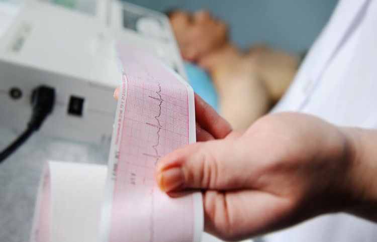
Kết quả đo điện tâm đồ giúp chẩn đoán thuyên tắc phổi
In addition, the electrocardiographic changes in the upper part are not unique to patients with pulmonary embolism. Such changes can occur in many acute or chronic cardiopulmonary diseases such as: Pneumonia, pneumothorax, recent pneumonectomy, upper airway obstruction, chronic obstructive pulmonary disease, cystic fibrosis , interstitial lung disease, severe kyphosis, sleep apnea,...
The fact that the electrocardiogram is not sensitive, is not specific enough to diagnose or rule out pulmonary embolism. Approximately 18% of patients with pulmonary embolism have a completely normal ECG. However, with compatible clinical features such as chest pain, hypoxemia, and electrocardiographic findings suggestive of pulmonary embolism, further testing is required to confirm the diagnosis.
Currently, at Vinmec International General Hospital, fibrinolysis is being applied for emergency treatment of patients with pulmonary embolism. With modern imaging equipment, CT perfusion results are available within 7 minutes and performed routinely. The technique is performed by a team of qualified, experienced doctors and modern equipment and professional services.
Master, Doctor Trinh Van Dong has nearly 10 years of experience in the specialty of Diagnostic Imaging, especially has strengths in performing techniques: X-ray, ultrasound, computed tomography, magnetic resonance. Currently, Dr. Dong is working and working at the Department of Diagnostic Imaging, Vinmec Ha Long International General Hospital.
To register for examination and treatment at Vinmec International General Hospital, you can contact Vinmec Health System nationwide, or register online HERE.
MORE :
Early signs of pulmonary embolism Complications of pulmonary embolism Risk of pulmonary embolism in pregnant and postpartum women


