This is an automatically translated article.
The article was professionally consulted with Master, Doctor Trinh Van Dong - Radiologist - Department of Diagnostic Imaging - Vinmec Ha Long International Hospital.Transfocal ultrasonography in neonates is an effective imaging technique for the diagnosis of a number of diseases. In particular, ultrasound through the fontanelle is very safe and does not affect the infant's body.
1. What is transfocal ultrasound?
Using the special anatomical characteristics of the newborn, experts have used the fontanel ultrasound method to diagnose the pathology of the central nervous system for children under 6 months of age, when the fontanelles have not yet closed. . Especially for premature babies, the risk of neuropathy is higher.The fontanelle is the incomplete position of the skull bone created by the skulls. When a baby is born, there are all 6 fontanels including:
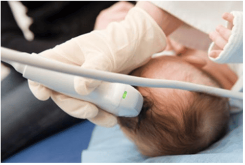
Anterior fontanelle: The largest in size, created between the two frontal bones and the two parietal bones. The average duration of closure was 14 months of age. Posterior fontanelle: Smaller than anterior fontanelle, made up of two parietal and occipital bones. Immediately after about 2-3 months old. The two temporal and two mastoid fontanelles are not talked about much, but they are also good ultrasound windows for the evaluation of neuropathy. Ultrasonography of the fontanelle uses an examination window including anterior fontanelle (this is a large window for good observation), posterior fontanelle, two temporal fonts and two mastoid fonts, which are good windows for evaluating the occipital lobe. Ultrasonography of the fontanelle is performed in children under 6 months of age when the skull bones are not fully developed and the skull joints have not yet healed. Ultrasound uses a single head due to the appropriate frequency to probe the child's fontanel area, the emitted ultrasound waves will capture images of brain structure. Brain structures including cerebral parenchyma, ventricles, thalamus, and blood vessels are visible on ultrasound of the fontanelle.
Advantages and disadvantages of transfocal ultrasound
Advantages:
Widely used, especially in premature infants. As an easy, low-cost method, ultrasound is almost non-invasive and can be done at the bedside. Ultrasonic waves are extremely safe and do not use radiation. Ultrasound scans give clear images of soft tissues that do not show up well on radiographs. Disadvantages:
When using ultrasound, it is difficult to evaluate the posterior fossa and convex surfaces of the brain. In term neonates with asphyxia within 24 h, no structural changes were observed on ultrasound. Difficult to assess lesions in pathology.
2. Indications for transfocal ultrasound
Fontanel ultrasound is indicated for the following subjects:For premature babies who need special care. For the subject, regular ultrasonography of the fontanelle is required to rule out neurological complications of preterm birth such as cerebral hemorrhage or periventricular white matter lesions due to asphyxia. Because the risk of this disease is higher in premature babies than in full-term babies. The child has an abnormal increase in head size. Bulging fontanelle Notice any abnormal neurological symptoms in the child. Like movement disorders, signs of meningoencephalitis syndrome appear ...
3. The role of transthoracic ultrasound
Transfocal ultrasound helps diagnose acquired neurological diseases and neurological abnormalities in children.
3.1 Acquired neuropathies detected by fontanel ultrasound Meningoencephalitis in infants at birth: fontanel ultrasound is very valuable in the diagnosis and prognosis of the disease. For preterm infants, the incidence of disease in preterm infants is up to 25-45%, low birth weight (<1500g) and less than 32 weeks, caused by increased pressure on blood vessels. For full-term infants, it is less common, accounting for 2-4%, caused by asphyxia, trauma, lack of clotting factors... Cerebellar hemorrhage: Common in premature infants, caused by anemia Brain; Birth trauma, blood clotting disorders... Using the fontanel ultrasound method, especially the ultrasound window is the posterior fontanelle, the temporal fontanelle to examine the occipital region. Support for the diagnosis of epidural hemorrhage: Rarely, due to strong trauma, often combined with computed tomography. Periventricular leukoplakia: The necrosis of the periventricular white matter due to ischemia, possibly due to fetal distress during pregnancy or birth. Ultrasound of the anterior fontanelle has a high diagnostic value. Detect tumors or cysts in the brain. Infectious diseases such as: sequelae of purulent meningitis, brain abscess... 3.2 Some congenital malformations detected by fontanal ultrasound Hydrocephalus: This is a dilated image of the lateral and third ventricles, with It can be caused by an imbalance between CSF production and absorption or by obstruction of outflow. Common causes are due to other malformations, pregnancy infection, intracranial tumor... Abnormalities of the corpus luteum: atrophy of the corpus callosum, lipoma of the male body Dandy-walker: An abnormality caused by the closing process of the tube caused by nerves. Common manifestations are hydrocephalus, hydrocephalus, and lobe aplasia. Water brain. Varicose veins of galen...
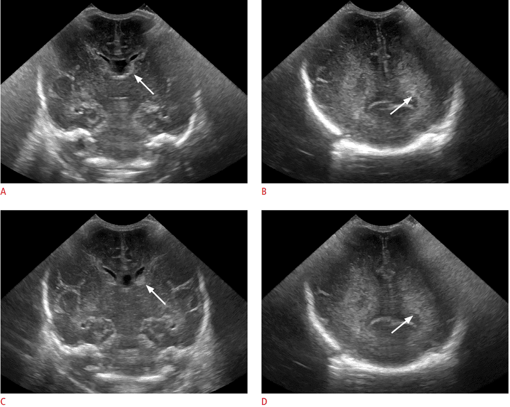
Ultrasonography of the fontanelle is of great value in the diagnosis of neonatal neuropathy. Especially this method is very simple, effective, low cost, safe but still very helpful in diagnosing diseases.
Infants who are born prematurely, have abnormalities in head size, bulging fontanelles or have neurological abnormalities should use transfocal ultrasound for timely diagnosis and treatment.
With a system of facilities, modern medical equipment, sterile space, minimizing the impact as well as the risk of disease spread, Vinmec will bring satisfaction to customers and receive benefits from customers. Highly appreciated by industry experts with:
Gathering a team of leading pediatricians: including leading experts with high professional qualifications (professor, associate professor, doctorate, master) , rich experience, worked at big hospitals like Bach Mai, 108.. The doctors are well-trained, professional, have a heart - reach, understand young psychology. In addition to domestic pediatric specialists, the Department of Pediatrics also has the participation of foreign experts (Japan, Singapore, Australia, USA) who are always pioneers in applying the latest and most effective treatment regimens. . Comprehensive services: In the field of Pediatrics, Vinmec provides a series of continuous medical examination and treatment services from Newborn to Pediatric and Vaccine,... according to international standards to help parents take care of their baby's health from birth to childhood. Advanced techniques: Vinmec has successfully deployed many specialized techniques to make the treatment of difficult diseases in pediatrics more effective: neurosurgery - skull, stem cell transplant blood in cancer treatment. Professional care: In addition to understanding children's psychology, Vinmec also pays special attention to the children's play space, helping them to play comfortably and get used to the hospital's environment, cooperate in treatment, improve the efficiency of medical treatment.
Please dial HOTLINE for more information or register for an appointment HERE. Download MyVinmec app to make appointments faster and to manage your bookings easily.


