This is an automatically translated article.
The article is professionally consulted by Master, Doctor Le Xuan Thiep - Radiologist - Department of Diagnostic Imaging - Vinmec Ha Long International Hospital.Renal artery MRI is a noninvasive technique that can reconstruct the anatomical image of the renal artery system and provide information about the physiological function of the kidney. Renal artery lesions can be observed without the use of contrast media.
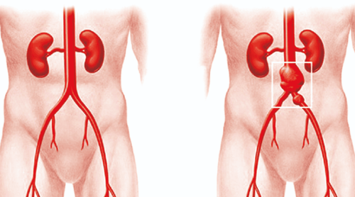
1. What is the renal artery?
The renal artery is a large branch of the abdominal aorta, entering at the renal hilum, which is responsible for supplying blood to the kidneys. The abdominal aorta gives branches to the renal artery at the level of the lumbar vertebral body L1-L2, below the bifurcation to the superior mesenteric artery. Each kidney is perfused by one renal artery (in some cases there may be 1-2 accessory renal arteries on one side). The right renal artery runs inferiorly and crosses posteriorly to the inferior vena cava and the right renal vein to reach the renal hilum. In contrast, the left renal artery is shorter and anterior than the right renal artery. The left renal artery is more horizontal, passing behind the left renal vein to enter the hilum, similar to the right renal artery. The renal artery has an average length of about 4 to 6 cm, with an average diameter of about 5 to 6 millimeters.Each renal artery divides into two large branches, dorsal and ventral, just anterior to the renal hilum. Then, they continue to divide many other small branches at the renal hilum before entering the parenchyma, including: posterior segment renal artery, lower segment renal artery, inferior anterior segment renal artery, segmental renal artery superior anterior lobe and superior segmental renal artery branch. These arterial branches then continue into the lobes and divide into many smaller branches, including the interlobular, interlobular, and arcuate branches. In addition, before dividing into dorsal and abdominal branches, the renal artery also gives many small arterial branches to neighboring organs such as the inferior adrenal artery and the ureteral artery. These branches are usually not visible on imaging studies.
2. What is renal artery MRI?
Renal artery MRI is a non-invasive technique that accurately evaluates renal artery anatomy and diagnoses causes of renal artery stenosis or occlusion with greater sensitivity and specificity than ultrasonography – especially especially in obese patients. Without the need for injections.Renal artery MRI helps to measure mean renal artery flow velocity, glomerular filtration rate and renal perfusion index. In addition, renal artery size is also accurately measured in renal angiography by magnetic resonance.
The biggest disadvantage of renal artery MRI is its high cost and contraindication in patients with pacemakers, metal clips, cochlear hearing aids, and some other prosthetic materials.

Bệnh nhân có thiết bị trợ thính ốc tai thì không được chụp MRI
3. When is an MRI of the renal artery indicated?
Indications for renal angiography are decided mainly in cases of suspected pathology related to renal artery abnormalities. Renal angiography has a role in diagnosis and monitoring of treatment, especially when other imaging modalities do not clearly examine abnormalities or the patient has contraindications to computed tomography of the renal artery. and contrast agents. Some diseases related to the renal artery system that require MRI are:Renal artery aneurysms Renal artery malformation Renal arteriovenous anastomosis Renal artery dissection Renal artery stenosis or occlusion due to fibrosis Atheroma, spasm Congenital renal artery atrophy Family history of dominant polycystic kidney disease Renal disease Fibromuscular dysplasia Chronic kidney disease hypertensive .
4. Contraindications to MRI of renal artery in which cases
Renal angiography is a safe, minimally invasive procedure and does not expose the patient to X-ray exposure. However, not all patients can undergo renal angiography. Renal artery MRI has its own contraindications, including:Patients who are carrying artificial implant materials in the body such as pacemakers, insulin injection chambers, artificial cochlea, artificial heart valves, etc. bone fusion equipment such as nails, screws clips for treating cerebral aneurysms shrapnel or bullets near the scan site Metallic objects in the eye Patients with claustrophobia Personal items such as claustrophobia jewelry, watches, phones, bank cards, computers, ...

Bệnh nhân có thiết bị điện tử như điện thoại chống chỉ định chụp MRI
5. Procedure for performing renal artery MRI
Renal artery MRI should be performed step by step according to the procedure to ensure patient safety and ensure image quality.Steps to perform renal artery MRI include:
Asking the patient's previous medical history, taking note of information related to surgery with implanted artificial materials to rule out contraindications . Ask the patient to remove personal items such as jewelry, phones, watches, and change clothes. This step should be performed with the patient's family in the room, if available. Explain and instruct the patient to follow the technician's requirements such as inhaling, exhaling, holding breath, ... Complete the procedures before conducting MRI of renal artery such as signing the commitment letter in case with contrast agent. When entering the imaging room, put the patient in the supine position, wrap the breathing wire around the patient's waist. Be careful not to wrap too tightly or too loosely. The coil is placed on the abdomen. Since the operation generates a lot of noise, the patient should wear noise-reducing headphones. When taking pictures, depending on the case, pulses are selected to combine with each other to record images of the renal artery system in turn. The basic pulses commonly used in renal artery MRI are axial 2D Fiesta pulses, axial T2 FRFSE pulses, coronal 2D Fiesta pulses, etc. The scan time lasts from 40 to 60 minutes. Vinmec International General Hospital with a system of modern facilities, medical equipment and a team of experts and doctors with many years of experience in neurological examination and treatment, patients can completely rest in peace. examination and treatment center at the Hospital.
Dr. Le Xuan Thiep has strengths in performing advanced and difficult magnetic resonance and computed tomography techniques such as: coronary computed tomography, cardiac function, cerebral magnetic resonance, perfusion brain and organs,..
If you notice any unusual health problems, you should visit and consult a specialist
Please dial HOTLINE for more information or register for an appointment HERE. Download MyVinmec app to make appointments faster and to manage your bookings easily.