This is an automatically translated article.
The article is expertly consulted by Master, Doctor Nguyen Thi Mai Anh - Doctor of Radiology - Department of Diagnostic Imaging and Nuclear Medicine - Vinmec Times City International General Hospital. Doctor Nguyen Thi Mai Anh has nearly 10 years of experience in the field of diagnostic imaging, especially in imaging breast and thyroid cancer.Preoperative localization of mammary gland parenchymal lesions with wire needles under the guidance of imaging tools including X-ray, ultrasound or magnetic resonance means. The purpose of the localization technique is to pinpoint the exact location of the tumor, and at the same time provide information about the "path" to the lesion from the skin for the surgeon to remove the lesion.
1. What is mammogram guided wire needle positioning?
Pinpoint localization of nonpalpable breast lesions and surgical removal of localized lesions for histopathology are the gold standard for breast cancer diagnosis.The determination of the lesion location is to measure the dimensions from the X-ray film, then the local needle will be poked. With the development of X-ray guided localization technique to increase positioning accuracy, breast lesions were compressed and fixed on the imaging table. This has the benefit of reducing the distance from the puncture site to the tumor and that the needle is positioned parallel to the chest wall. After that, the development of ultrasound-guided localization technique has been increasingly developed and brought many good results.
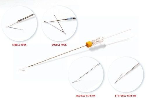
Kim dây định vị u vú Sinurep
In conditions where mammography-guided core needle biopsies are not available, wire needle localization and open biopsy are used for the diagnosis of suspected nonpalpable lesions. In cases where the lesion has been identified on pathologic core needle biopsies, the role of localization and surgical excision is complete resection of the lesion (atypical hyperplasia of breast tissue, surgical scars, lobular breast cancer in situ, ductal carcinoma in situ, invasive cancer with small size and no extraparenchymal invasion...). In addition, in cases where there is no compatibility between histopathology and core needle biopsy, this technique is also indicated to confirm the diagnosis because of the large number of specimens and can help achieve the goal of treatment. good treatment.
2. Purpose of locating the wire needle under the guidance of mammogram
In the pre-intervention practice of patients with abnormal lesions in the breast parenchyma, the surgeon can only see the patient's skin in the operating room, so it is not possible to find non-palpable lesions or microscopic organisms. Suspicious calcifications detected on ultrasound or radiographs. The main purpose is to guide the surgeon, the radiologist has inserted the wire needle to locate the center or near the lesion, then the surgeon will follow the locating needle to remove the lesion. love.Today, with the advancement of breast ultrasound and mammography, suspicious lesions can be detected even when the size is very small and the patient cannot feel it, so it is necessary to In order to obtain the lesion for treatment, diagnose the pathology and with minimal surgical goal but achieve maximum efficiency, this preoperative needle positioning technique plays an increasingly important role. In particular, positioning under X-ray guidance to remove lesions, especially microcalcifications, is one of the early detection signs of breast cancer that has been used for a long time, before the tools were available. ultrasound guide. Since then, the technique of locating mammary gland lesions under X-ray guidance has created many advantages for surgeons when approaching patients at the location of the lesion.

Hình ảnh máy X-quang vú Mammomat
3. How to perform the technique of locating mammary gland lesions under X-ray guidance
3.1. Preparing instruments
Positioning needle: wire needle with hook and wire needle for breast positioning wire with diameter of positioning needle 20G, needle core 25G, needle length 10cm, wire core 15cm Positioning media: X-ray machine, breast tissue press with many holes with lead mark locator Prepare anesthetic, sterile supplies, locating needle and shockproof case3.2. Prepare the patient
The patient was explained, wrote a commitment and changed the surgical gown. Sterilized with Betadin alcohol at the breast skin on the lesion.
Bệnh nhân cần được sát trùng bằng cồn Betadin trước khi thực hiện
3.3. How to proceed?
X-ray by pressing forceps with hole first position (use 90 degree tilt or straight up and down first depending on lesion location), mark the position of lesion visible on film against skin when breast is still pressed. Re-sterilize the marked position, insert the needle positioned vertically into the lesion just enough depth while the breast is still pressed. Take the film again to confirm that the needle is positioned correctly at the lesion site and the needle direction is vertical (the tip of the needle covers the tip of the needle). Slowly release the presser, avoid shifting the patient too much because it may cause the needle to move, guide the patient to stand out of the machine gently. Continue to take pictures in the orthogonal plane to determine the distance from the locating needle to the lesion, insert the locating needle marked with the cm ruler on the needle sheath to the lesion. Take a picture to confirm that the locating needle has reached the lesion, remove the needle cover, leave the needle core, mark a small lead on the skin where the needle is inserted, take an X-ray film to determine the position of the wire needle with a plane integrity. Fix the needle on the outer part of the skin. Draw a location map. Save the image of the patient sample when the surgeon dissects the lesion and locates the wire, and informs the surgeon. Mark the lesion on the specimen and send it to pathology.
Tư thế chụp X quang vú
4. Complications after performing the technique of locating mammary gland lesions under X-ray guidance
Complications of radiographically guided mammography are uncommon. Botanically, this is a very safe technique if performed correctly and surgically. Serious complications such as bleeding, pneumothorax (due to the needle going too deep into the chest wall) are very rare but need to be treated quickly and promptly.The more common complication is the vagus nerve reaction because the patient is often anxious, nervous, painful, afraid, maybe even fainting... interrupting the surgery. Therefore, the initial consultation and interpretation of the patient is very important. In addition, there are possible side effects that are severe pain and prolonged bleeding at the needle insertion site... but can be improved by squeezing the breast or using pain relievers.
In addition, there are complications that occur when the locating needle is not removed, the needle tip will migrate to other locations, the chest wall muscle, the pleural cavity, the base of the neck, and may even migrate down to the base of the neck. butt. To limit the loss of the locator cord, the positioning technique needs to be accurate, the surgical technique must be good, and the coordination of mapping between the radiologist and the surgeon must be good.
In summary, mammographic-guided wire needle placement is a safe, low-complication technique that aims to orient the surgeon with minimal intervention for maximum efficiency in one stroke. Because of this purpose, the technique is increasingly being applied widely, becoming an effective means of locating, especially in the case of small, hard-to-touch tumors, suspected of breast cancer.

Khách hàng có thể đến Bệnh viện Đa khoa Quốc tế Vinmec để được tầm soát ung thư vú
Examination and consultation with an oncologist. Breast cancer screening with bilateral breast ultrasound and mammogram. Up to now, Vinmec has become a prestigious address in breast cancer screening with:
Team of highly qualified and experienced doctors. Comprehensive professional cooperation with domestic and international hospitals: Singapore, Japan, USA, etc. Comprehensive treatment and care for patients, multi-specialty coordination towards individualizing each patient. There is a full range of specialized facilities to diagnose the disease and stage it before treatment: Endoscopy, CT scan, PET-CT scan, MRI, histopathological diagnosis, genetic test - cytology, Biopsy ... There are full range of mainstream cancer treatment methods: surgery, radiation therapy, chemotherapy, stem cell transplant...
Please dial HOTLINE for more information or register for an appointment HERE. Download MyVinmec app to make appointments faster and to manage your bookings easily.
MORE:
9 risk factors for breast cancer in women When should a mammogram be performed? What is the significance of breast ultrasound in breast cancer screening?