This is an automatically translated article.
The article is professionally consulted by MSc, BS. Dang Manh Cuong - Doctor of Radiology - Department of Diagnostic Imaging - Vinmec Central Park International General Hospital. The doctor has over 18 years of experience in the field of ultrasound - diagnostic imaging.Magnetic resonance imaging (MRI) of the face and neck with magnetic contrast injection is similar to the non-injection technique to help examine the anatomical structures of the face and neck. However, thanks to the contrast-enhancing effect of magnetic contrast agents, it will help increase the ability to evaluate inflammatory lesions, tumor lesions, especially beneficial in cases where vascular and perfusion properties are required. . So what is the procedure of magnetic resonance imaging with injection of magnetic contrast?
1. Outline
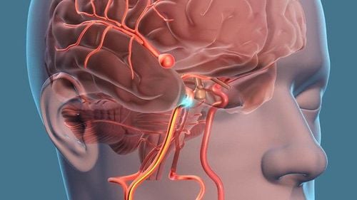
The face-neck area is always a challenge for doctors because there are many complex anatomical structures here. The vascular system that supplies blood to the facial sensory organs and the major vessels in the neck that supply blood to the brain are the targets of contrast-enhanced magnetic resonance imaging (MRI).Besides examining the bone and soft tissue structures in the face and neck area, it is similar to MRI without contrast. Using contrast agent from injection into blood vessels helps to increase contrast and easily detect vascular anatomy in the face - neck area.
Thanks to image reconstruction algorithms, the doctor can visualize the path of blood vessels, detect abnormalities, blood damage in the face - neck area. In addition, knowing the perfusion time from the artery to the capillary at the target organ and back to the vein, magnetic resonance imaging can assess the perfusion nature of the organ or the extent of vascular proliferation of the lesion. u.
2. Indications for magnetic resonance imaging with magnetic contrast injection
Injuries to the face - neck; Inflammatory lesions, infections of the face - neck; Assess size, location, density and perfusion of tumor lesions; Investigation of vascular abnormalities in the face and neck; Some other indications depend on professional treatment requirements.
Đối tượng bị chấn thương vùng mặt – cổ cần được chụp cộng hưởng từ
3. Contraindications to magnetic resonance imaging with injection of magnetic contrast agents
3.1. Absolute contraindications Similar contraindications for non-contrast magnetic resonance imaging such as: implants of metal-based electronic devices such as pacemakers, cochlear electrodes, injection devices continuously under the skin; metal foreign bodies in the body; Intracranial, orbital, vascular metal implants < 6 months. Allergy or hypersensitivity to contrast agents. 3.2. Relative contraindications In addition to relative contraindications such as magnetic resonance imaging without injection, there are: metal implants > 6 months, agoraphobia, claustrophobia. Relative contraindications for contrast agents + Glomerular filtration rate less than 30 ml/min+ Pregnant and lactating women
4. Facial-neck magnetic resonance imaging procedure with injection of magnetic contrast agent
4.1 Preparation before proceeding Performer: radiologist and radiologist specialized in imaging; Equipment: 1.5 Tesla or larger resonator; Magnetic Contrast Drugs; Shockproof box; Other drugs: Sedative support drugs can be used in cases where the patient has fear of the dark, claustrophobia; Patient: can have a light meal, it is not necessary to fast before the scan. Instructions and explanations of what to do before filming begins. Patient removes contraindicated items.
Hệ thống máy chụp cộng hưởng từ 1.5 Tesla

Kết quả phim cộng hưởng từ giúp bác sĩ đưa ra hướng điều trị phù hợp và chính xác nhất cho người bệnh
5. Follow-up during and after magnetic resonance imaging
During magnetic resonance imaging: closely monitor vital signs (pulse, blood pressure, breathing rate), consciousness and focal neurological signs of the patient. After injection, closely monitor vital signs and blood pressure to prevent allergies and anaphylaxis to magnetic contrast agents.6. Complications and treatment directions
Some patients with agoraphobia, loneliness or claustrophobia may be fearful and agitated. The technician or doctor needs to guide and encourage the patient to comfort or use sedatives in case of indication. Complications related to contrast agents need to be treated and diagnosed quickly according to the anaphylaxis protocol. Vinmec International General Hospital is currently one of the major hospitals with modern machinery and equipment for general medical examination and treatment and accurate and modern MRI for brain and vascular diseases. brain in particular.Especially, Vinmec International General Hospital is the first unit in Southeast Asia to put into use the new 3.0 Tesla Silent Resonance Imaging machine from the US manufacturer GE Healthcare.
The machine currently applies the safest and most accurate magnetic resonance imaging technology available today, without using X-rays, non-invasive. Silent technology is very beneficial for patients who are young children, the elderly, patients with weak health or have just had surgery.
Please dial HOTLINE for more information or register for an appointment HERE. Download MyVinmec app to make appointments faster and to manage your bookings easily.