This is an automatically translated article.
The article is professionally consulted by Dr. Nguyen Dinh Hung - Diagnostic Imaging - Department of Diagnostic Imaging - Vinmec Hai Phong International General Hospital.
The liver is one of the most important organs and is considered the energy factory for the body. With conventional diagnostic techniques, magnetic resonance imaging can also diagnose and monitor lesions in the liver and biliary tract. However, in order to investigate more deeply and approach the physical damage of the liver and distinguish it from some other diseases, it is necessary to take liver magnetic resonance imaging with tissue-specific contrast agents. Therefore, the liver is the only abdominal organ that has its own specific magnetic resonance imaging procedure.
1. Outline
The liver is a solid organ located in the upper abdomen, tilted to the right, has the main task of metabolizing and synthesizing substances for the body as well as secreting bile to participate in the digestive process. In Vietnam, liver malignancies such as liver cancer, cholangiocarcinoma have a high rate and are the leading cause of death. Therefore, the correct diagnosis helps to give the right and timely treatment direction, improving the patient's prognosis.
The vascular system of the liver from many sources and histology also has many differences with other organs, so for the liver, to help increase the diagnostic ability, it is necessary to use a liver-specific contrast agent. Currently, Primovist is a tissue-specific magnetic contrast agent, used in liver magnetic resonance imaging.
Primovist is a paramagnetic contrast agent, strongly soluble in water and absorbed by hepatocytes about 50% of the dose, excreted by the biliary tract (50%) and the kidney (50%). This allows the degree of hepatic perfusion to be examined and the morphological assessment of the biliary tract. In addition, on the T1W pulse sequence, the drug shortens the relaxation time, so it increases the signal and increases the contrast image in the liver tissue.
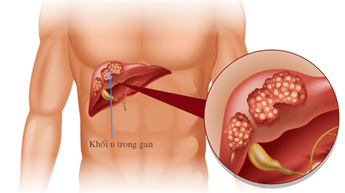
Chụp cộng hưởng từ gan giúp chẩn đoán ung thư gan
2. Advantages of liver magnetic resonance with liver tissue-specific contrast agent
With one injection brings two benefits. One is to help investigate the extracellular and vascular properties in the kinetic phase similar to that of contrast agents from conventional Gadolinium. In addition, Primovist drug also helps to study hepatobiliary properties in the late phase 10 - 20 minutes (50% biliary and 50% renal). Hepatocellular carcinoma such as primary hepatocellular carcinoma (HCC), does not contain Kupffer cells and normal hepatocytes, does not respond to late-stage therapy to increase sensitivity and specificity.
In addition, Primovist helps to differentiate adenomas (hepatocellular tumors), focal nodular hyperplasia (FNH), detect metastases, cholangiocarcinomas and partially assess liver function. Increase the rate of early detection of primary liver cancer, which is safe, easy to perform and increases the survival rate for patients
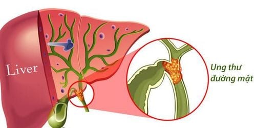
Chụp cộng hưởng từ gan có thuốc đối quang từ đặc hiệu mô giúp phát hiện ung thư đường mật
3. Indications for liver magnetic resonance imaging with tissue-specific magnetic contrast injection.
Suspected liver damage after initial imaging methods; ● Distinguishing malignant lesions in the liver from other pathological causes;
● Assess stage, extent of invasion and metastasis of liver cancer;
● Investigation of biliary tract morphology in bile duct diseases.
4. Contraindication to magnetic resonance imaging with injection of magnetic contrast agent.
Absolutely contraindicated in patients who carry metal-based electromagnetic devices such as pacemakers, anti-vibration machines, cochlear electrode implants, automatic subcutaneous injection devices. Surgical implantation of intracranial means, blood vessels, eye sockets < 6 months, the patient is in critical condition. ● Relative contraindications for patients carrying metal surgical instruments > 6 months, agoraphobia, fear of the dark, glomerular filtration rate < 30 mL/min, pregnant or lactating women .
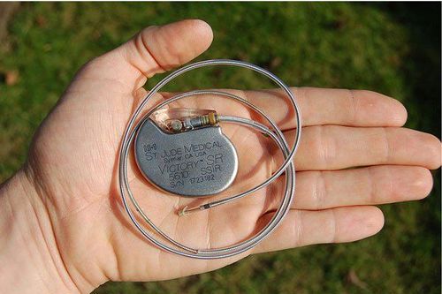
Bệnh nhân có mang máy tạo nhịp chống chỉ định tuyệt đối chụp cộng hưởng gan
5. Liver magnetic resonance imaging procedures with tissue-specific drugs
Step 1: The patient is explained how to breathe, tries to cooperate, prepare what to do, and remove contraindicated items.
● Step 2: Take localization, take before injection with a layer thickness of 5-8 mm, choose commonly used pulse sequences such as SE, STIR T2W in cross-section and horizontal, T1W before drug injection.
● Step 3: Inject contrast agent from 2ml/sec with 0.05 mmol/kg body weight and perform post-injection dynamic study in arterial phase, portal vein phase, equilibrium phase and late phase after 20 minutes .
● Step 4: The copper and phase opposite GRE pulse sequence for fat detection is examined before injection.
● Step 5: The technician prints the film or saves the CD, transfers the data to the doctor's workstation to read the results.
In addition, there is now a diffusion technique (DWI) applied in the assessment of cancer staging and response to treatment.
Tiêm thuốc đối quang trong chụp MRI gan từ 2ml/giây với liệu 0.05 mmol/kg cân nặng
7. Read the results of liver magnetic resonance imaging with tissue-specific contrast agents
Obtained images clearly show liver anatomy and biliary structures. ● Detecting lesions, if any, as well as assessing the extent of drug enhancement at the lesion site.
Vinmec International General Hospital is currently one of the major hospitals with modern machinery and equipment for general medical examination and treatment procedures and accurate and modern MRI for brain and vascular diseases. brain blood in particular.
Especially, Vinmec International General Hospital is the first unit in Southeast Asia to put into use the new 3.0 Tesla Silent Resonance Imaging machine from the US manufacturer GE Healthcare.
The machine currently applies the safest and most accurate magnetic resonance imaging technology available today, without using X-rays, non-invasive. Silent technology is very beneficial for patients who are young children, the elderly, patients with weak health or have just had surgery.
Doctor Nguyen Dinh Hung has over 10 years of experience in the field of diagnostic imaging (Ultrasound, CT, MRI). Trained and practiced on hepatobiliary interventional radiology at Bach Mai Hospital (Intervention under ultrasound guidance, DSA, CT...) and deployed at the Diagnostic Imaging Department of Viet Tiep Hospital Hai Phong. Currently, he is a doctor at the Diagnostic Imaging Department of Vinmec Hai Phong International General Hospital.
Please dial HOTLINE for more information or register for an appointment HERE. Download MyVinmec app to make appointments faster and to manage your bookings easily.