This is an automatically translated article.
This article was professionally consulted by a Doctor of Radiology, Department of Radiology - Vinmec Central Park International General Hospital.Doppler ultrasound is one of the diagnostic tools with high value, especially in diseases of the heart and blood vessels. However, for accurate and highly effective image analysis, understanding the principle of pulse Doppler ultrasound also plays an extremely important role.
1.Principle of Doppler vascular ultrasound
Doppler ultrasound creates images based on basic principles:
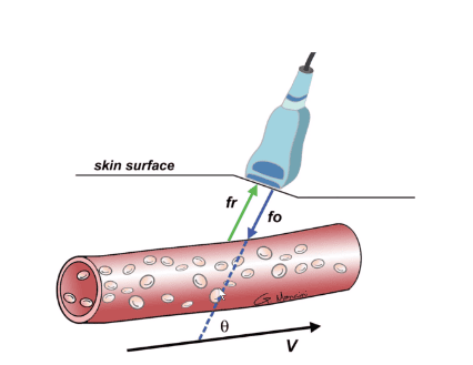
Using the frequency change of sound waves, electromagnetic waves when the source moves relative to the observer. For example, objects must be in motion to apply the Doppler effect (such as blood vessel flow, urine, cardiac hemodynamics, pleural motion survey, etc.). For other organs, if vascular assessment is not required, only 2D ultrasound is sufficient. The probe will emit a frequency (f0) that will match the direction of velocity (v) of the red blood cells by an angle θ (Doppler angle). When this wave reaches the red blood cells, it will bounce back as a reflected wave (fr). When red blood cells (blood vessels) are stationary: f0 and fr are equal. When the blood flow is moving then certainly f0 and fr are different depending on the direction of flow.

Siêu âm Doppler mạch máu
In fact, when Doppler ultrasound of the pulse:
Doppler angle < 60 degrees will have an accurate measurement (the smaller the better). Manual scanning, changing the scanning angle will change the Doppler angle. The “steer” mode can reduce the Doppler angle (will be useful in examining peripheral blood vessels). After the wave is reflected, the Doppler effect will be displayed in different forms:
Flow velocity displayed as sound (eg echocardiogram) Coded as color. Encode as Doppler spectrum.
2. Survey value of each Doppler ultrasound system
Doppler ultrasound includes types: continuous Doppler, pulse Doppler, fast Doppler, energy Doppler, Duplex, Triplex...and has great advantages in evaluating heart and blood vessel pathology.
2.1. Continuous Doppler (CW) Ultrasound Continuity Doppler ultrasound probe consists of 2 crystals: one side emits ultrasound waves, the other receives the reflected beam. The process of transmitting and receiving Doppler waves takes place continuously. To calculate the speed of the object moving, average the frequencies of the emitted and received beams.
The advantage of continuous Doppler ultrasound is that it can measure very large blood flow velocities, which is most suitable for echocardiography. The downside is that the speed cannot be recorded at a specified point.
2.2. Pulsed Doppler Ultrasound (PW) The pulse Doppler ultrasound probe has only one crystal: it emits intermittent sound waves, pauses for a while, receives the echo, and then resumes.
The advantage of pulsed Doppler ultrasound is the recording of blood flow velocity at a defined location. In the form of Doppler spectrum can calculate many hemodynamic parameters, so pulse Doppler is often used for vascular ultrasound.
On the contrary, this type of ultrasound also has disadvantages: limiting the maximum speed that can be measured, the latter limit can be probed as well as highly dependent on the Doppler angle.
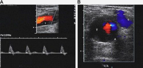
2.3. Color Doppler Ultrasound (CDI) Color Doppler ultrasound is essentially pulsed Doppler but takes information from many points (multiple ports). Color Doppler will have many imaging lines forming lines, on each line there are many gates. The number of lines and ports are encapsulated in a single cross-sectional area called a color box.

Siêu âm Doppler màu (CDI)
The signal from these reflected waves is coded as a composite color into a flow color map with the following characteristics:
Flow direction: the closer you get to the probe, the red color, the more color you go away from the probe green. The magnitude of the velocity is expressed through the intensity of the color: the darker the color, the greater the speed, the lighter the smaller the speed. The advantage of color Doppler ultrasound is to record the flow direction on the anatomical background, location, and size of the blood flow.
The disadvantage is that the hemodynamic parameters of the blood flow cannot be evaluated, it is easy to interfere, and it exceeds the threshold. Therefore, in order to be able to fully analyze the morphology and hemodynamic properties of blood vessels, it is recommended to combine 2-D ultrasound, color Doppler image and Doppler spectrum.
2.4. Energy Doppler Ultrasound Energy Doppler ultrasound displays an ultrasound image with only red (or additional yellow-orange) color as the dominant color. Power Doppler only answers the presence or absence of vascular flow, helping to assess perfusion.
The advantages of energy Doppler ultrasound are high sensitivity, low noise, low velocity flow detection, no over-threshold phenomenon. The downside is that it's prone to motion artifacts.
3.Application of Doppler ultrasound

Siêu âm Doppler mạch máu
Ultrasound of blood vessels is a very valuable imaging method, not only to help evaluate vascular lesions but also to show hemodynamic changes, thereby helping doctors make treatment decisions. suitable treatment.
Objectives of Doppler ultrasound:
Detect blood flow. Determine the direction of the flow. Determine flow pattern: arterial-venous, normal or abnormal. Measure the flow rate. Practical applications of different types of Doppler ultrasound:
In cardiac survey: applying continuous Doppler ultrasound. In vascular examination: Duplex or Triplex ultrasound In the study of tumor perfusion, organ perfusion: apply color Doppler or power Doppler (preferred because of its high sensitivity). In fetal circulation survey: assess fetal heart, ductus venosus, umbilical artery, middle cerebral artery, uterine artery. In general, each Doppler system has its own advantages and disadvantages and requires flexible application. The sonographer must master the principles of vascular Doppler ultrasound, ultrasound techniques and have more knowledge of pathology to adjust the appropriate parameters.
Vinmec International General Hospital is one of the hospitals that not only ensures professional quality with a team of leading medical doctors, modern equipment and technology, but also stands out for its examination and consultation services. comprehensive and professional medical consultation and treatment; civilized, polite, safe and sterilizing medical space. on the website for service.
Please dial HOTLINE for more information or register for an appointment HERE. Download MyVinmec app to make appointments faster and to manage your bookings easily.


