This is an automatically translated article.
Pleural effusion is a common infection in diseases of the respiratory system. In the early stages, it can be successfully treated with antibiotics combined with aspiration or drainage of the pleura. If not diagnosed in time, it will form pleural deposits and leave serious sequelae.1. What is pleural deposits?
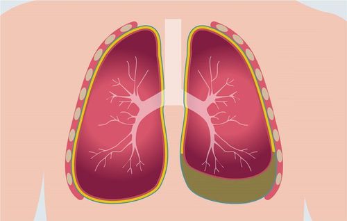
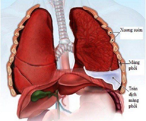
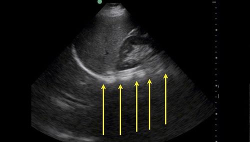
Pleural deposits is a pathological condition consisting of two main types of lesions: first, there is an actual space between the lung and the chest wall; In other cases, the outer surface of the lung is covered by a layer of fibrous tissue that binds so that the lung cannot expand. Pleural deposits may form 3 to 5 weeks after the onset of pleurisy.
1.1 Indications for treatment of pleural deposits
Cases of pleural deposits are determined by clinical and subclinical. Patients are indicated for surgery according to the anatomical stage, whereby the earlier the surgery, the simpler and more effective the better.
The main surgical technique is to peel off the visceral pleura and clean the pleural cavity. When there is a severe infection of the pleura, it is necessary to add local treatment techniques and high-dose, broad-spectrum systemic antibiotics and drug combinations.
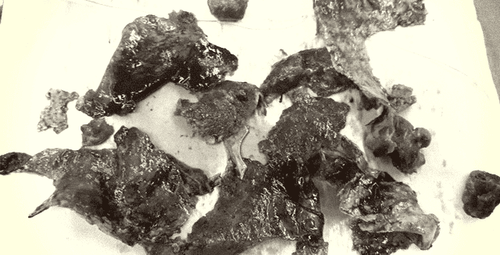
Ổ cặn mủ màng phổi mạn tính
1.2 Contraindications to treatment of pleural deposits
Existing foci of infection of other organs, severe chronic disease, contralateral lung injury that does not allow ventilation of one lung...
2. Surgical procedure to peel the pleura
2.1 Implementation staff: 2 teams
Surgical team: The main doctor must be a cardiothoracic and thoracic surgeon, 2 assistants, 1 instrument and 1 running outside of cardiology. The vascular and thoracic anesthesiology team includes an anesthesiologist and 1-2 assistants.
2.2 Means
2.2.1 Surgical instruments
Chest opener and closure kit, major surgical instrument for common thoracic surgery. Some other tools are specific to lung surgery.2.2.2 Anesthesia means
Preparation kit for thoracic surgery and drugs for anesthesia and cardiovascular resuscitation. Double-bore endotracheal tube (Carlens)...2.2.3 Patients
Prepare for surgery according to the procedure of heart and chest surgery (especially hygiene, prophylactic antibiotics). Anesthesia resuscitation examination. Medical staff need to explain the patient and family as prescribed. Finalize legal documents. Before entering surgery, the patient and family are clearly explained before surgery about the disease status; Complications, complications, and sequelae in unfortunate cases may be caused by disease, surgery, anesthesia, anesthesia, pain relief, or the patient's physical condition. Stable treatment of other medical diseases that the patient may have such as: High blood pressure, diabetes, ... before surgical intervention (except for emergency surgery). The patient needs a blood transfusion if the anemia is severe. The patient may or may not receive prophylactic antibiotics before surgery.
2.2.4 Medical records
The doctor completes the medical records according to the general regulations of breast surgery (ultrasound, tests, x-rays ...) and checks all legal procedures including minutes of consultation, stamps ....2.3 Steps to take
Check records: Fully comply with regulations (administrative, professional, legal). Checking the patient: Confirming the right person (name, age ...), correct disease. The patient is told to lie on his side 900 to the opposite side, with a pillow across the chest. Perform a posterior-lateral thoracotomy through the V intercostal space into the pleural space, creating a surgical field into the pleural space because the lung is very sticky (care must be taken to avoid injuring the healthy lung parenchyma) Identify the lesion and evaluate the entire damage to the remaining lobes of the lung, the lymphatic system, the pleura... Anesthetize and prepare the patient: Double-bore endotracheal anesthesia; Monitor electrocardiogram and capillary oxygen saturation (SpO2) continuously.
2.4 Technical
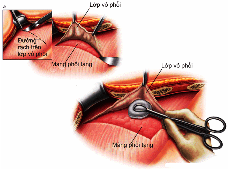
Các bước tiến hành phẫu thuật bóc vỏ màng phổi là gì?
Step 1: Start dissection to remove adhesions, approach the pleural deposits (it is recommended to use an electric knife to avoid bleeding from the areas of lung adhesion). Step 2: The patient is cleaned of the pleural cavity, especially the location of the deposits. Specimens of the removed pleural deposits were sent for pathology and microbiology. Step 3: In case the patient has a long-term pleural deposit that causes thickening of the pleura (the surface of the lung parenchyma is covered with a thick layer of organized fibrin), it is necessary to remove this layer as much as possible to help the lungs expand. . Step 4: Check for air leaks at positions on the lung parenchyma where the organized fibrin layer is removed, use sterile serum to pour into the pleural cavity and inflate the lung to check (if air leaks appear). across the surface of the lung parenchyma, requiring immediate suturing). Step 5: The doctor performs hemostasis, irrigates the chest and places two silicone drains into the pleural cavity (anterior and posterior) and continuously drains and vacuums immediately after to prevent occlusion due to blood clots. Step 6: Close the chest after good lung expansion.
2.5 Tracking
Patients should be tested for blood gases, electrolytes, liver and kidney function, blood count, hematocrit immediately after returning to the recovery room for 15-30 minutes. Bed chest X-ray (best). Perform hemodynamics, respiration, drainage, urine every 30 minutes - 1 hour, in the first 24 hours or longer depending on hemodynamic status. Doctors prescribe antibiotics to prevent infections, heart drugs, diuretics, pain relievers; blood transfusion and blood replacement solutions ... depending on the patient's condition. Respiratory therapy in the first day after surgery.
2.6 Handling of accidents
Postoperative bleeding: The patient is indicated for emergency hemostasis if bleeding > 100ml/hour + hemodynamic disturbances; or > 200ml/hour for 3 consecutive hours. Post-operative atelectasis is usually caused by the patient's poor breathing and extensive sputum obstruction after surgery. Clinical examination of patients with dyspnea, fever, reduced alveolar murmur; X-ray showed atelectasis. Besides, it is necessary to provide good pain relief for the patient, systemic antibiotics, the patient needs to sit up early, shake and cough up sputum. If necessary, bronchoscopy can be performed. Any questions that need to be answered by a specialist doctor as well as if you have a need for medical examination and treatment at Vinmec International General Hospital, you can contact Vinmec Health System nationwide or register online HERE.
MORE:
Causes and diagnosis of pneumothorax Is pleural effusion dangerous? Diagnostic test for pleural effusion


