This is an automatically translated article.
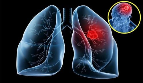
The article is professionally consulted by doctors working at Oncology Center - Vinmec Central Park International General HospitalLung cancer is the most common malignancy, accounting for about one-third of all cancer deaths, and it is the leading cause of cancer-related death in both men and women. Although rates of lung cancer for men are falling, rates for women continue to rise. Even more worrisome is the increased incidence of non-small cell lung cancer (NSCLC) in patients who have never smoked or have a minimal smoking history (eg, < 10 to 15 packs). / five).
1. Causes of lung cancer
The first is smoking. Smoking is the cause of 85% to 90% of lung cancer cases; The risk of lung cancer in smokers is 30 times higher than in non-smokers. Passive smoking can double your risk of lung cancer. Second, asbestos dust is related to the cause of malignant mesothelioma. Asbestos exposure also increases the risk of lung cancer, especially in smokers (the risk is three times higher than with smoking alone). In addition, radiation exposure may increase the risk of small cell lung cancer in both smokers and nonsmokers. Radon rays are associated with 6% of lung cancer cases. Other substances linked to lung cancer include arsenic, nickel, chromium compounds, chloromethyl ether and air pollutants.
Currently, there are two main types of lung cancer: Non-small cell lung cancer (NSCLC). Small cell lung cancer (SCLC)
The most common type of lung cancer is NSCLC. NSCLC accounts for 8.5 out of 10 of all lung cancers diagnosed. NSCLC can be divided into subtypes by looking at the cancer cells under a microscope. This is called histological classification.
There are 3 histological types of NSCLC: First, adenocarcinoma occurs in the cells lining the alveoli (small air sacs) in the lungs. This is the most common type. About 4 out of 10 lung cancers are adenocarcinomas.
Second, squamous cell carcinoma (squamous) occurs in the flat cells that line the inside of the airways in the lungs. About 3 out of 10 lung cancers are squamous cell carcinomas. Third, large cell (undifferentiated) carcinomas that start in large cells can develop anywhere in the lung. This accounts for 1.5 out of 10 lung cancers.

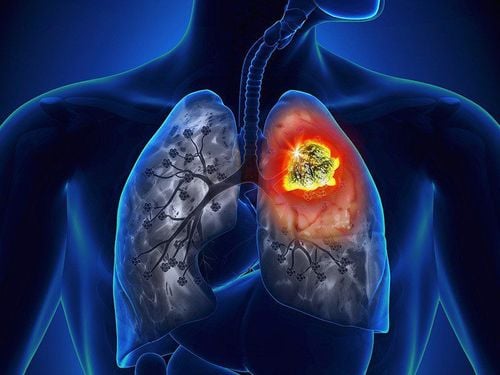
Hút thuốc lá là nguyên nhân của 85% đến 90% trường hợp ung thư phổi
2. Symptoms and signs
Symptom . Most patients are symptomatic. Lung cancer symptoms may be due to primary disease in the chest (dry cough, hoarseness, hemoptysis, chest pain, dyspnea, pneumonia), metastatic disease (new lymph node mass, bone pain, pathological fractures) , headache, convulsions), or a paraneoplastic syndrome (loss of appetite, weight loss, nausea due to hypercalcemia, etc.).
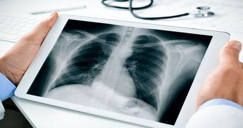
However, patients can also be completely asymptomatic and incidentally detected on chest x-ray or chest CT. Patients with cancer located at the top of the lung (Pancoast tumor) may experience paresthesias and weakness in the arms and hands as well as Horner's syndrome (ptosis, hyperhidrosis, and pupillary constriction) due to compression. of a tumor involving the cervical sympathetic nerves. Evidence of metastatic disease includes bone pain (bone metastases); jaundice and abdominal symptoms with rapidly enlarged liver (liver metastases) or enlarged lymph nodes.
Laboratory signs: Computed tomography (CT) scan of the chest and abdomen. Chest CT for lung cancer staging is clearly superior to chest radiographs and has been reported to have an overall accuracy of 70%. Mediastinal lymph nodes are usually considered abnormal when greater than 1.5 cm in diameter and normal when less than 1.0 cm. Chest CT scan also provides information on the extent of invasion of the primary tumor, the presence of pleural effusion, and lymph node status.
Adrenal tumor pictures. Adrenal metastases are very common in NSCLC. It is sometimes possible to distinguish between metastatic disease and an adrenal adenoma based on densification features on CT or MRI. If the diagnosis is equivocal and the adrenal gland is the only site of suspected metastasis, biopsy is required to clarify the diagnosis.
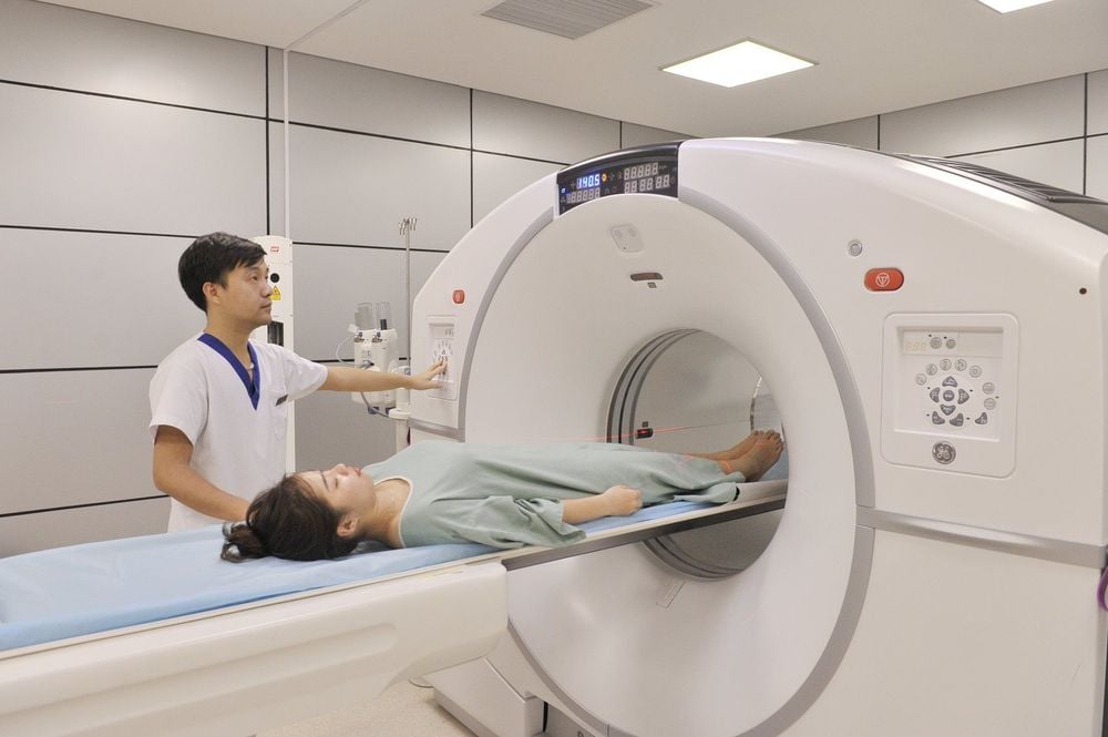
Chụp cắt lớp CT tại Bệnh viện Đa khoa Quốc tế Vinmec
3. Lung Biopsy
Recently, cytological examination of sputum samples has been largely replaced by flexible bronchoscopy. Even under the most thorough investigations, repeated sputum cytology is positive in only 60% to 80% of centrally located tumors and 15% to 20% of central tumors. Peripheral.
Bronchoscopy (bronchoscopy) with a flexible bronchoscope if there is symptomatic or radiographic evidence of an accessible mass. Most cancers can be diagnosed by direct screening. Occasionally, tumors that are evident only with extrinsic bronchial stenosis can be diagnosed through bronchoscopy with transbronchial biopsies. Bronchoscopy is not necessary if a histological or cytological diagnosis of metastatic lung cancer has been made. Endoscopic bronchial ultrasound (EBUS) and GPS coordinate bronchoscopy significantly improve bronchoscopy's ability to sample mediastinal lymph nodes and evaluate further lesions.
Lymph nodes. Peripheral lymph nodes in the supraclavicular or axillary regions represent another potential site for biopsy.
4. Stage Evaluation
After a histological diagnosis of lung cancer, the evaluation of lung cancer should focus on determining whether the disease is confined to the chest region and can therefore be treated with a curative intent (SCLC). localized stage and NSCLC stages I to III) or whether the patient has advanced disease.
Positron emission tomography (PET-CT). Although the PET-CT scan has demonstrated superiority over CT scanning and complements mediastinoscopy in the evaluation of mediastinal nodes, it is most useful in excluding distant metastases. PET may also be useful in assessing response after preoperative treatment (that is, chemotherapy) or in post-treatment follow-up. PET-CT scanners are now in common use and further improve patients' ability to accurately stage.
Brain CT or brain MRI is recommended as part of staging for patients with SCLC, which is associated with a 10% incidence of brain metastases without neurological symptoms. These tests are not recommended for most patients with small tumor stage I NSCLC in the absence of clinical signs. All patients with more advanced NSCLC and all patients with neurological symptoms should have brain scans.
Percutaneous and transthoracic needle biopsies are often used to diagnose lung cancer in cases where the tumor is located near the skin. Bone scintigraphy has largely been replaced by PET-CT. However, bone scintigraphy provides additional information regarding bone disease for PET-CT and is less expensive.
5. Genetic testing and immunohistochemistry in stage IV . patients
Patients with NSCLC need to be investigated for important gene mutations because about 60% of NSCLC stage IV cases in Vietnam have EGFR mutations. These cases are indicated for treatment with drugs that target EGFR inhibitors. In addition, patients also need to investigate other gene mutations such as ROS1, MET to serve as a basis for targeted drug therapy.
In addition, cases without gene mutations need immunohistochemistry for PD-L1 factor, which is the basis for the treatment of immune checkpoint inhibitors.
Oncology Center - Vinmec Central Park International General Hospital is modernly built according to international standards, using a multi-specialty approach model in diagnosis and selection of appropriate treatment regimens for each patient. contribute to comprehensive patient care. Vinmec Central Park Oncology Center has outstanding advantages in cancer treatment such as:
Vinmec Converging experts in radiation, chemotherapy and surgery, equipped with modern facilities. The most modern Truebeam radiotherapy planning system and machine in Southeast Asia, with the outstanding advantage of minimizing the effects of radiation on benign tissues compared to X-rays, reducing irradiation time and the risk of adverse effects. side effects for patients.) helps effectively treat common cancers: Lung, Head, neck, breast.... As the first hospital in Ho Chi Minh City to apply autologous immunotherapy, Vinmec Hospital Central Park can treat cancer with multimodal cancer treatment regimens suitable for each case, bringing high treatment efficiency, providing one more reliable cancer examination and treatment address for people. people in Ho Chi Minh City and surrounding areas, so that people do not have to go abroad for treatment According to Saigon Investment.


