This is an automatically translated article.
The article was professionally consulted by Specialist Doctor I Tran Cong Trinh - Radiologist - Radiology Department - Vinmec Central Park International General Hospital. The doctor has many years of experience in the field of diagnostic imaging.1. Outline
The pelvic region in men and women is different because of the different anatomical structures of the genitals. The female pelvis contains the uterine organs, two bilateral appendages, the cervix, and the vagina, and in the male contains the prostate gland. The organs of the urinary system such as bladder, ureters, ureters; Digestive system such as rectum, anus are relatively similar.When a suspected pelvic lesion is detected after initial imaging, magnetic resonance imaging may be indicated in these cases for the purpose of surveying the pelvic organ and lesion.
Pelvic magnetic resonance imaging provides clear, high-resolution and good-contrast images that improve patient diagnosis and prognosis in treatment.
2. Indications for pelvic magnetic resonance imaging without injecting magnetic contrast
Diagnosis and staging of malignant tumors in the vagina, cervix, uterus, ovaries and fallopian tubes; Differentiate tumor or inflammation of adnexa, abscess of adnexa. Pain due to suspected endometriosis or uterine leiomyomas; Detection and identification of congenital abnormalities in the male and female pelvis. In the case of uterine fibroids, the patient may be assigned to take a scan when the surgeon needs to survey the number and location of the tumor before surgery, hysterectomy or embolism; Monitor and evaluate for recurrence of bowel, bladder and prostate tumors or gynecological tumors after surgical treatment; Diagnosis of causes of abdominal pain in pregnant women includes appendicitis and abnormal masses in the uterus and ovaries.
3. Contraindications to pelvic magnetic resonance imaging without contrast injection
Absolutely contraindicated in patients with a history of implantation of metal-based electronic devices such as pacemakers, defibrillators, cochlear electrodes, metal surgical facilities less than 6 months, critical medical conditions . Relative contraindications: patients with claustrophobia, fear of solitude, fear of the dark, surgical tools with metal over 6 months.4. Preparation for pelvic magnetic resonance imaging without contrast injection
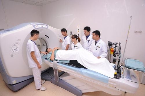
Medical staff including radiologists and technicians; Vehicles with resonance imaging machines of 1.5 Tesla or more; Sedative; Patients need to coordinate well with the doctor during the scan and remove contraindicated items as required.5. Steps to conduct pelvic magnetic resonance imaging without injecting magnetic contrast
5.1 Preparation before taking the picture With the patient in the supine position, the technician selects the position of the receiver coil and moves the table into the magnetic field of the machine and locates the imaging area.5.2. Selection of pulse sequences for non-contrast pelvic magnetic resonance imaging from the initial localization; Pulse sequence 1: Horizontal T2W with fat removal, slice thickness 6cm, interval between slices 10% of slice thickness (0.6 mm or coefficient 1.0-1.1), L-R code scale,, FOV < 200mm , the cross-sectional saturation plate is placed above the slice to prevent vascular interference; horizontal plane against abdominal wall fat interference. Pulse sequence 2: T1W cross-sectional plane, fat removal, position of slices similar to sequence 1. Pulse sequence 3: T2W with fat removal, horizontal plane or subframe position, thickness 4-6 mm, spaced 10 % of the thickness of the cut, the plate is saturated in the horizontal plane against vascular interference. Pulse sequence 4: T2W in vertical plane, thickness 5mm, interval 10% of cut thickness (0 – 0.5mm). Note: it is possible to combine drugs to help reduce intestinal motility, instruct the patient to breathe to reduce the noise caused by breathing.
6. Read results of pelvic magnetic resonance imaging without contrast injection
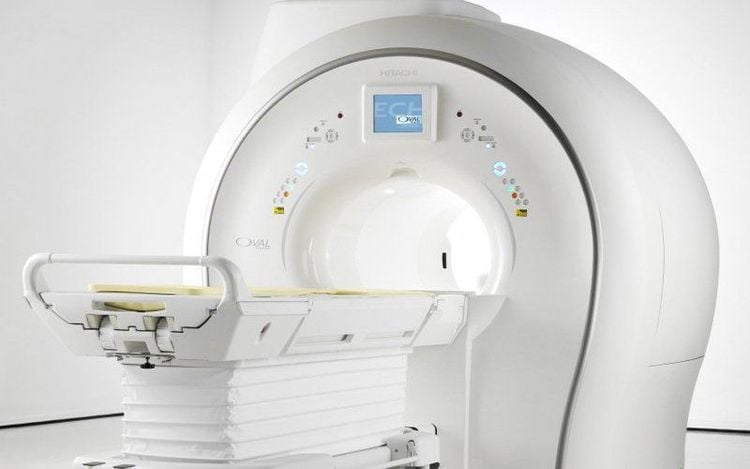
Especially, Vinmec International General Hospital is the first unit in Southeast Asia to put into use the new 3.0 Tesla Silent Resonance Imaging machine from the US manufacturer GE Healthcare.
The machine currently applies the safest and most accurate magnetic resonance imaging technology available today, without using X-rays, non-invasive. Silent technology is very beneficial for patients who are young children, the elderly, patients with weak health or have just had surgery.
Please dial HOTLINE for more information or register for an appointment HERE. Download MyVinmec app to make appointments faster and to manage your bookings easily.


