This is an automatically translated article.
The article is professionally consulted by Master, Resident Doctor Nguyen Van Anh - Radiologist - Department of Diagnostic Imaging and Nuclear Medicine - Vinmec Times City International General Hospital.When the patient has signs of jaundice, itchy hands and feet, pain in the epigastrium or is being treated for pancreatic diseases... pancreatic ultrasound will be the indicated technique. This is a simple and effective method to help doctors survey the condition of the pancreas as well as the response to treatment of pancreatic diseases.
1. What is Pancreatic Ultrasound?
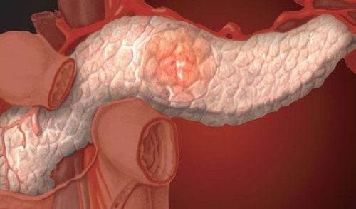
The pancreas is an organ located deep in the abdomen, just in front of the spine, behind the peritoneum. The pancreas is about 15 cm long, about 6 cm high and about 3 cm thick and weighs about 80 g, with two main functions: (1) The exocrine function of producing and excreting pancreatic juices (containing enzymes) helps for the process of digesting food; (2) Endocrine function helps regulate sugar metabolism and a number of other related processes.Previously, to survey the condition of the pancreas, doctors often used X-ray methods, so only indirectly surveying the pancreas, with limited information. Today, with the development of technology, ultrasound of the pancreas has become an important part of the investigation of pancreatic conditions and pathologies.
Although ultrasound of the pancreas is an effective method to help doctors identify pancreatic diseases, there are some difficulties that can be encountered during an ultrasound such as the pancreas, such as: retroperitoneal position, small size, and frequent abnormalities of the pancreas. masked by gas in the stomach and colon, especially in people who may be obese.

Vị trí cấu tạo của tụy trong cơ thể
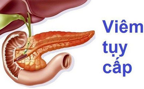
2. In which cases need an ultrasound of the pancreas?
In the following cases, the doctor will appoint the patient to do an ultrasound of the pancreas:● The patient has jaundice, itching, suspected of having liver disease.
Patient presents with epigastric pain, vomiting of unknown cause
Patient has rapid weight loss
Persistent diarrhea
Left pleural effusion
Alcoholic patients often have pancreatic diseases
Patients with persistent abdominal pain
Patients with abdominal trauma due to traffic accidents, work accidents or other accidents
Monitor complications of acute pancreatitis and disease progression through treatment
● Patients with suspected pancreatic cancer .
3. Pancreatic ultrasound technique
3.1 What preparation should be done before an ultrasound of the pancreas? ● Before the pancreatic ultrasound, the patient should fast for 6-8 hours, preferably in the morning (without breakfast), food will be digested after waking up and an empty stomach will help image. more accurate ultrasound.
Người bệnh nên nhịn ăn trước khi siêu âm để giúp kết quả siêu âm chính xác hơn
● Wearing loose, comfortable clothes when going for an ultrasound will make the examination process easier.
● In case of emergency ultrasound, ultrasound can be performed directly without fasting.
3.2 Technical process of pancreatic ultrasound The following are the steps to perform a standard pancreatic ultrasound:
First of all, information about the patient's name, age, and other information will be compared with the records and medical records .
● Patient's position: Lie on your back, arms raised above your head, legs straight. Exposing the abdomen from the tip of the breastbone to the pubic joint, relax naturally.
● Ask about the patient's symptoms and medical history.
● Apply gel to the abdomen
● During the ultrasound, several positions can be used to better reveal the image of the pancreas such as: lying on the right side, left side or stomach, or in combination with Breathe, breathe according to the rhythm of the doctor's instructions.

Uống nhiều nước trong quá trình siêu âm giúp cho hình ảnh rõ ràng hơn
4. Limitations and errors in pancreatic ultrasound
Pancreatic ultrasound can be done in most people. However, in some of the following cases, pancreatic ultrasound may bring limited results, need to be combined with some other additional techniques:● Patient is too fat
● Patient has paralytic ileus, distention
● Newly operated or laparoscopy patient
● Baryte gastrointestinal scan patient...
In addition, some diseases can be confused with pancreatic lesions on ultrasound such as:
● The patient has a solid retroperitoneal tumor, especially an adrenal tumor
● The retroperitoneal lymph node.
Colonic waste
● Confuse dilated Wirsung duct with splenic vein.
Master, Resident Doctor Nguyen Van Anh graduated as a Resident Doctor in Diagnostic Imaging at Hanoi Medical University and attended training in Teaching and Scientific Research Competency at the University General Sydney, Australia, internship at Concord Hospital Radiology Department, Sydney, Australia. Dr. Van Anh has strengths in magnetic resonance imaging, computed tomography, X-ray, ultrasound; specialized in musculoskeletal imaging; tumor biopsies (bone, lung, liver...) under the guidance of computed tomography and ultrasound; aspiration cytology (lymph nodes, thyroid, breast,...); Drain the abscesses under ultrasound guidance. Any questions that need to be answered by a specialist doctor as well as if you want to be examined and treated at Vinmec International General Hospital, you can contact Vinmec Health System nationwide or register online. online HERE.
MORE
Role of the pancreas in the endocrine system Abdominal ultrasound is an ultrasound of which parts? What can an abdominal ultrasound reveal?